Homologous 2',5'-phosphodiesterases from disparate RNA viruses antagonize antiviral innate immunity
- PMID: 23878220
- PMCID: PMC3740845
- DOI: 10.1073/pnas.1306917110
Homologous 2',5'-phosphodiesterases from disparate RNA viruses antagonize antiviral innate immunity
Abstract
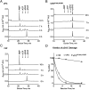
Efficient and productive virus infection often requires viral countermeasures that block innate immunity. The IFN-inducible 2',5'-oligoadenylate (2-5A) synthetases (OASs) and ribonuclease (RNase) L are components of a potent host antiviral pathway. We previously showed that murine coronavirus (MHV) accessory protein ns2, a 2H phosphoesterase superfamily member, is a phosphodiesterase (PDE) that cleaves 2-5A, thereby preventing activation of RNase L. The PDE activity of ns2 is required for MHV replication in macrophages and for hepatitis. Here, we show that group A rotavirus (RVA), an important cause of acute gastroenteritis in children worldwide, encodes a similar PDE. The RVA PDE forms the carboxy-terminal domain of the minor core protein VP3 (VP3-CTD) and shares sequence and predicted structural homology with ns2, including two catalytic HxT/S motifs. Bacterially expressed VP3-CTD exhibited 2',5'-PDE activity, which cleaved 2-5A in vitro. In addition, VP3-CTD expressed transiently in mammalian cells depleted 2-5A levels induced by OAS activation with poly(rI):poly(rC), preventing RNase L activation. In the context of recombinant chimeric MHV expressing inactive ns2, VP3-CTD restored the ability of the virus to replicate efficiently in macrophages or in the livers of infected mice, whereas mutant viruses expressing inactive VP3-CTD (H718A or H798R) were attenuated. In addition, chimeric viruses expressing either active ns2 or VP3-CTD, but not nonfunctional equivalents, were able to protect ribosomal RNA from RNase L-mediated degradation. Thus, VP3-CTD is a 2',5'-PDE able to functionally substitute for ns2 in MHV infection. Remarkably, therefore, two disparate RNA viruses encode proteins with homologous 2',5'-PDEs that antagonize activation of innate immunity.
Keywords: Nidovirales; RNA capping enzyme; Reoviridae; interferon-stiumulated gene.
Conflict of interest statement
The authors declare no conflict of interest.
Figures


Similar articles
-
Lineage A Betacoronavirus NS2 Proteins and the Homologous Torovirus Berne pp1a Carboxy-Terminal Domain Are Phosphodiesterases That Antagonize Activation of RNase L.J Virol. 2017 Feb 14;91(5):e02201-16. doi: 10.1128/JVI.02201-16. Print 2017 Mar 1. J Virol. 2017. PMID: 28003490 Free PMC article.
-
Murine AKAP7 has a 2',5'-phosphodiesterase domain that can complement an inactive murine coronavirus ns2 gene.mBio. 2014 Jul 1;5(4):e01312-14. doi: 10.1128/mBio.01312-14. mBio. 2014. PMID: 24987090 Free PMC article.
-
Rotavirus Controls Activation of the 2'-5'-Oligoadenylate Synthetase/RNase L Pathway Using at Least Two Distinct Mechanisms.J Virol. 2015 Dec;89(23):12145-53. doi: 10.1128/JVI.01874-15. Epub 2015 Sep 23. J Virol. 2015. PMID: 26401041 Free PMC article.
-
Viral phosphodiesterases that antagonize double-stranded RNA signaling to RNase L by degrading 2-5A.J Interferon Cytokine Res. 2014 Jun;34(6):455-63. doi: 10.1089/jir.2014.0007. J Interferon Cytokine Res. 2014. PMID: 24905202 Free PMC article. Review.
-
Silencing the alarms: Innate immune antagonism by rotavirus NSP1 and VP3.Virology. 2015 May;479-480:75-84. doi: 10.1016/j.virol.2015.01.006. Epub 2015 Feb 25. Virology. 2015. PMID: 25724417 Free PMC article. Review.
Cited by
-
Birth of protein folds and functions in the virome.Nature. 2024 Sep;633(8030):710-717. doi: 10.1038/s41586-024-07809-y. Epub 2024 Aug 26. Nature. 2024. PMID: 39187718 Free PMC article.
-
Ribonuclease L and metal-ion-independent endoribonuclease cleavage sites in host and viral RNAs.Nucleic Acids Res. 2014 Apr;42(8):5202-16. doi: 10.1093/nar/gku118. Epub 2014 Feb 5. Nucleic Acids Res. 2014. PMID: 24500209 Free PMC article.
-
Lineage A Betacoronavirus NS2 Proteins and the Homologous Torovirus Berne pp1a Carboxy-Terminal Domain Are Phosphodiesterases That Antagonize Activation of RNase L.J Virol. 2017 Feb 14;91(5):e02201-16. doi: 10.1128/JVI.02201-16. Print 2017 Mar 1. J Virol. 2017. PMID: 28003490 Free PMC article.
-
The Rotavirus Interferon Antagonist NSP1: Many Targets, Many Questions.J Virol. 2016 May 12;90(11):5212-5215. doi: 10.1128/JVI.03068-15. Print 2016 Jun 1. J Virol. 2016. PMID: 27009959 Free PMC article. Review.
-
ABCE1 Regulates RNase L-Induced Autophagy during Viral Infections.Viruses. 2021 Feb 18;13(2):315. doi: 10.3390/v13020315. Viruses. 2021. PMID: 33670646 Free PMC article.
References
-
- Dong B, Silverman RH. 2-5A-dependent RNase molecules dimerize during activation by 2-5A. J Biol Chem. 1995;270(8):4133–4137. - PubMed
-
- de Groot RJ, et al. Family Coronaviridae. In: King AMQ, Adams MJ, Carstens EB, Lefkowitz EJ, editors. Virus Taxonomy, Ninth Report of the International Committee on Taxonomy of Viruses. Amsterdam: Elsevier; 2012. pp. 806–828.
Publication types
MeSH terms
Substances
Grants and funding
LinkOut - more resources
Full Text Sources
Other Literature Sources
Molecular Biology Databases

