. 2013 Aug 6;110(32):13186-91.
doi: 10.1073/pnas.1300910110. Epub 2013 Jul 22.

Ca2+ release-activated Ca2+ channel blockade as a potential tool in antipancreatitis therapy

Affiliations

Ca2+ release-activated Ca2+ channel blockade as a potential tool in antipancreatitis therapy

Julia V Gerasimenko et al. Proc Natl Acad Sci U S A. .

Abstract

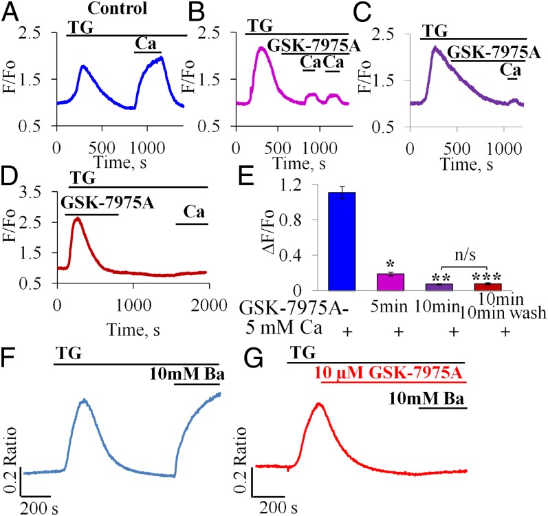
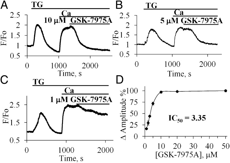
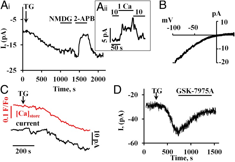
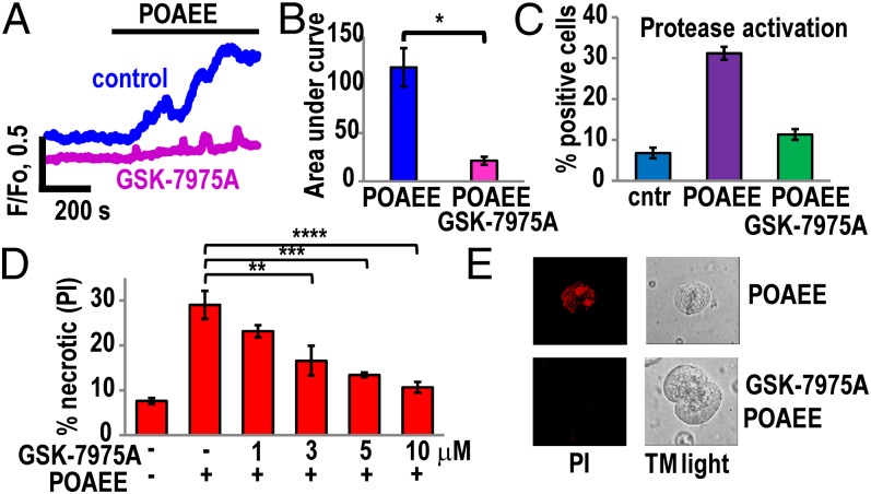
Alcohol-related acute pancreatitis can be mediated by a combination of alcohol and fatty acids (fatty acid ethyl esters) and is initiated by a sustained elevation of the Ca(2+) concentration inside pancreatic acinar cells ([Ca(2+)]i), due to excessive release of Ca(2+) stored inside the cells followed by Ca(2+) entry from the interstitial fluid. The sustained [Ca(2+)]i elevation activates intracellular digestive proenzymes resulting in necrosis and inflammation. We tested the hypothesis that pharmacological blockade of store-operated or Ca(2+) release-activated Ca(2+) channels (CRAC) would prevent sustained elevation of [Ca(2+)]i and therefore protease activation and necrosis. In isolated mouse pancreatic acinar cells, CRAC channels were activated by blocking Ca(2+) ATPase pumps in the endoplasmic reticulum with thapsigargin in the absence of external Ca(2+). Ca(2+) entry then occurred upon admission of Ca(2+) to the extracellular solution. The CRAC channel blocker developed by GlaxoSmithKline, GSK-7975A, inhibited store-operated Ca(2+) entry in a concentration-dependent manner within the range of 1 to 50 μM (IC50 = 3.4 μM), but had little or no effect on the physiological Ca(2+) spiking evoked by acetylcholine or cholecystokinin. Palmitoleic acid ethyl ester (100 μM), an important mediator of alcohol-related pancreatitis, evoked a sustained elevation of [Ca(2+)]i, which was markedly reduced by CRAC blockade. Importantly, the palmitoleic acid ethyl ester-induced trypsin and protease activity as well as necrosis were almost abolished by blocking CRAC channels. There is currently no specific treatment of pancreatitis, but our data show that pharmacological CRAC blockade is highly effective against toxic [Ca(2+)]i elevation, necrosis, and trypsin/protease activity and therefore has potential to effectively treat pancreatitis.

Keywords: AR42J; alcohol metabolite; capacitative Ca2+ entry; hepatocyte Ca2+ entry; pancreas.


Conflict of interest statement

The authors declare no conflict of interest.

Figures

Fig. 1.
Fig. 1.
CRAC channel blocker inhibits Ca2+ and Ba2+ influx. (A–C) Effects of different periods of incubation [5 min, n = 5 (B) and 10 min, n = 14 (C)] of pancreatic acinar cells with GSK-7975A (10 μM) compared with control (A, n = 5) (15 min incubation did not significantly further increase the degree of blockade, n = 3). (D) Washout of GSK-7975A did not result in recovery of the Ca2+ signal within ∼10 min. (E) Summary of results shown in AD. Mean [Ca2+]i amplitude change (ΔF/F0) due to Ca2+ influx (n = 5, example trace in A) was dramatically reduced after 5 min with GSK-7975A (n = 14, *P < 10−6, example trace in B) and reduced further after 10 min with GSK-7975A (n = 14, **P < 10−11, example in C). Inhibition was effectively irreversible (after washing out for 10 min) as amplitude remained very low (n = 7, ***P < 10−8, example trace in D). As seen in E, there was no significant difference in averaged amplitudes from experiments of the type shown in C and D (P > 0.69); whereas the averaged amplitudes from the type of experiments shown in B and C are significantly different (P < 10−6). Data presented as mean ± SEM (F and G) GSK-7975A inhibits Ba2+ influx in pancreatic acinar cells. Representative traces of changes in [Ba2+]i (using Fura-2) due to Ba2+ influx in cells exposed to GSK-7975A (10 μM) for 10 min (G) compared with control untreated cells (F).
Fig. 2.
Fig. 2.
Concentration dependence of the inhibitory effect of GSK-7975A on the elevated [Ca2+]i following readmission of external Ca2+ after thapsigargin treatment. (A–C) Effects of acute application of GSK-7975A in different concentrations [10 μM (A), 5 μM (B), 1 μM (C)] on the elevated [Ca2+]i plateau in the presence of 5 mM CaCl2. (D) Summary of the results of the experiments on the concentration dependence of the inhibitory effect of GSK-7975A normalized to the inhibitory effect of 50 μM (100%).
Fig. 3.
Fig. 3.
Store-operated ionic currents, developing after thapsigargin treatment, recorded with the whole cell patch clamp configuration. The individual traces shown were all recorded at a holding potential of −50 mV and with an external [Ca2+] of 10 mM. To prevent activation of the large Ca2+-dependent ion currents in acinar cells (13), patch clamp pipettes were filled with a solution containing a mixture of 10 mM BAPTA and 2 mM Ca2+. (A, i) Inward current induced by bath application of 2 μM TG to empty the ER Ca2+ content. Replacing Na+ with NMDG+ had little effect on the inward current, but 100 μM of 2-APB practically abolished the current. This effect was rapidly reversible. (A, ii) Reducing the external Ca2+ concentration from 10 to 1 mM (replacement of CaCl2 by MgCl2) reduced reversibly the stable maximal plateau amplitude of the inward current during TG exposure. (B) Representative I/V curve obtained using a voltage ramp protocol (0.4 V/s) from −100 mV to 40 mV (difference between ramp registration before and after 2-APB application). (C) Simultaneous measurements of changes in the intrastore [Ca2+] and the membrane current following TG application. The Upper red trace shows the gradual reduction of the intrastore Ca2+ concentration recorded by changes of Fluo-5N fluorescence. The Lower black trace shows the development of the inward current. (D) GSK-7975 (10 μM) inhibits markedly the inward current evoked by application of 2 μM TG.
Fig. 4.
Fig. 4.
GSK-7975A dramatically reduces Ca2+ overload and necrosis induced by the fatty acid ethyl ester POAEE. (A) Representative traces of changes in [Ca2+]i in response to 100 μM POAEE in the absence (n = 8) and presence of GSK-7975A (10 μM) (n = 12). (B) Quantitative analysis of experiments of the type shown in A by comparing the integrated [Ca2+]i elevation above the baseline (area under the curve) recorded during the first 10 min of POAEE application. Blue bar represents the control (without GSK-7975A), whereas the red bar represents the test results (cells pretreated with 10 μM GSK-7975A for 10 min before application of POAEE). The mean values (±SEM) are significantly different, P < 0.0002. (C) Pretreatment of cells with 10 μM GSK-7975A for 10 min inhibited by 64% (green bar) protease activation induced by POAEE (100 μM) (purple bar) as measured with generic protease substrate bis-l-aspartic acid amide rhodamine 110 (D2-R110). Blue bar represents the results from control (not exposed to POAEE) cells. The mean values (±SEM) in control and GSK-7975A plus POAEE treatment are not significantly different, P = 0.05; n = 4, >200 cells in each group. (D) POAEE (100 μM)-induced necrosis was dramatically reduced in cells treated with 1 μM, 3 μM, 5 μM, and 10 μM GSK-7975A for 10 min. In the control series of experiments (no POAEE treatment), the level of necrosis was low (n = 3 series of experiments with number of tested cells in each group >350). Inhibition of necrosis was significant for 3 μM GSK-7975A (**P < 0.02) and highly significant for 5 and 10 μM GSK-7975A (***P < 0.003 and ****P < 0.001). (E) Necrosis was visualized by staining cells with propidium iodide (PI). All experiments were performed in the presence of 1 mM CaCl2.

References

    1. Pandol SJ, Saluja AK, Imrie CW, Banks PA. Acute pancreatitis: Bench to the bedside. Gastroenterology. 2007;132(3):1127–1151. - PubMed
    1. Petersen OH, Sutton R. Ca2+ signalling and pancreatitis: Effects of alcohol, bile and coffee. Trends Pharmacol Sci. 2006;27(2):113–120. - PubMed
    1. Hegyi P, Pandol S, Venglovecz V, Rakonczay Z., Jr The acinar-ductal tango in the pathogenesis of acute pancreatitis. Gut. 2011;60(4):544–552. - PMC - PubMed
    1. Petersen OH, et al. Fatty acids, alcohol and fatty acid ethyl esters: Toxic Ca2+ signal generation and pancreatitis. Cell Calcium. 2009;45(6):634–642. - PubMed
    1. Petersen OH, Tepikin AV. Polarized calcium signaling in exocrine gland cells. Annu Rev Physiol. 2008;70:273–299. - PubMed

Publication types

MeSH terms