. 2013 Jul 22;202(2):261-76.
doi: 10.1083/jcb.201304181.

Chromatin insulator bodies are nuclear structures that form in response to osmotic stress and cell death

Affiliations

Chromatin insulator bodies are nuclear structures that form in response to osmotic stress and cell death

Todd Schoborg et al. J Cell Biol. .

Abstract

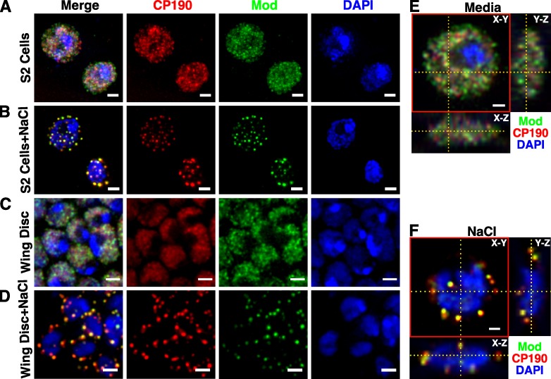
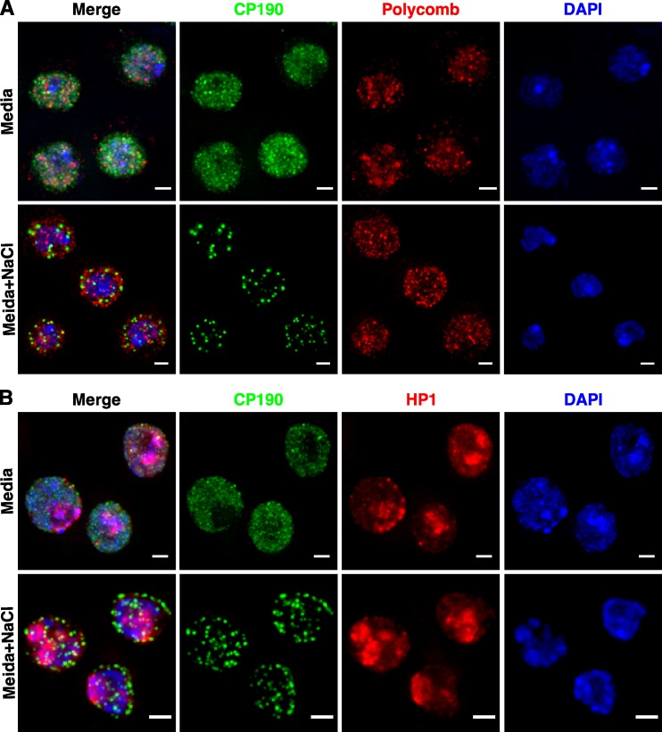
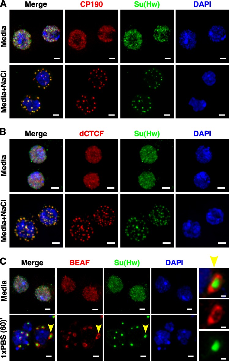
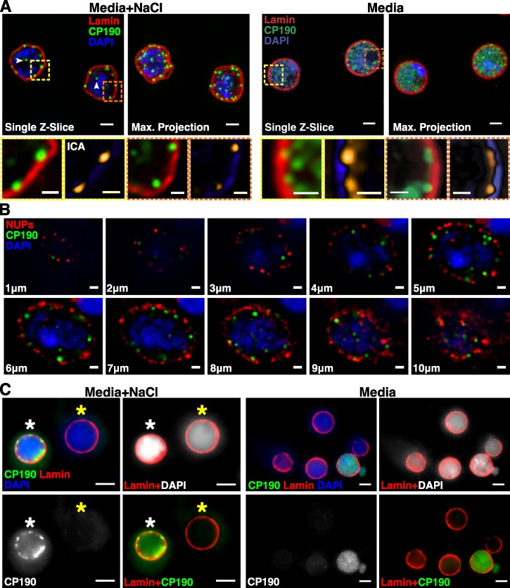
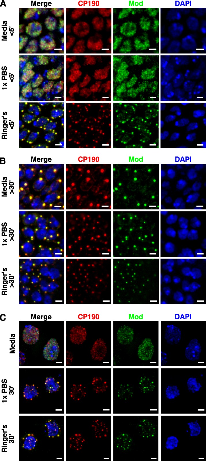
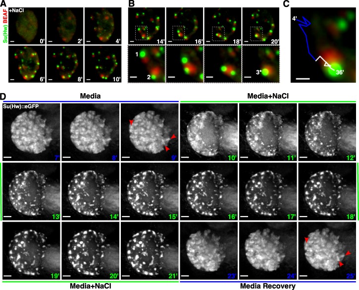
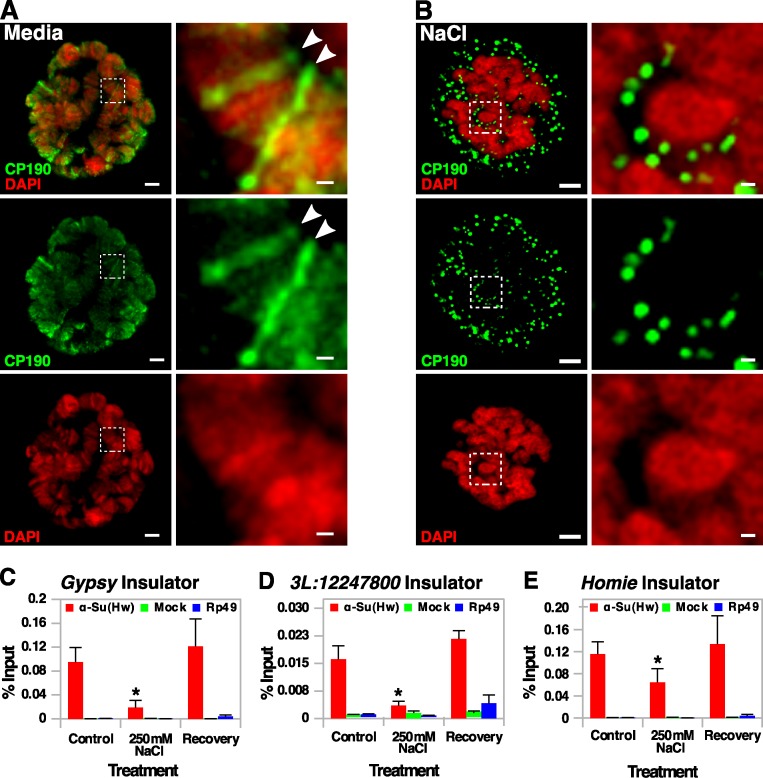
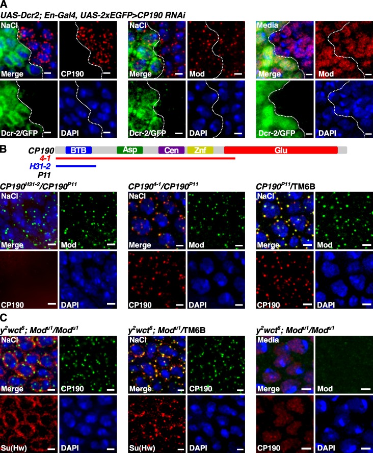
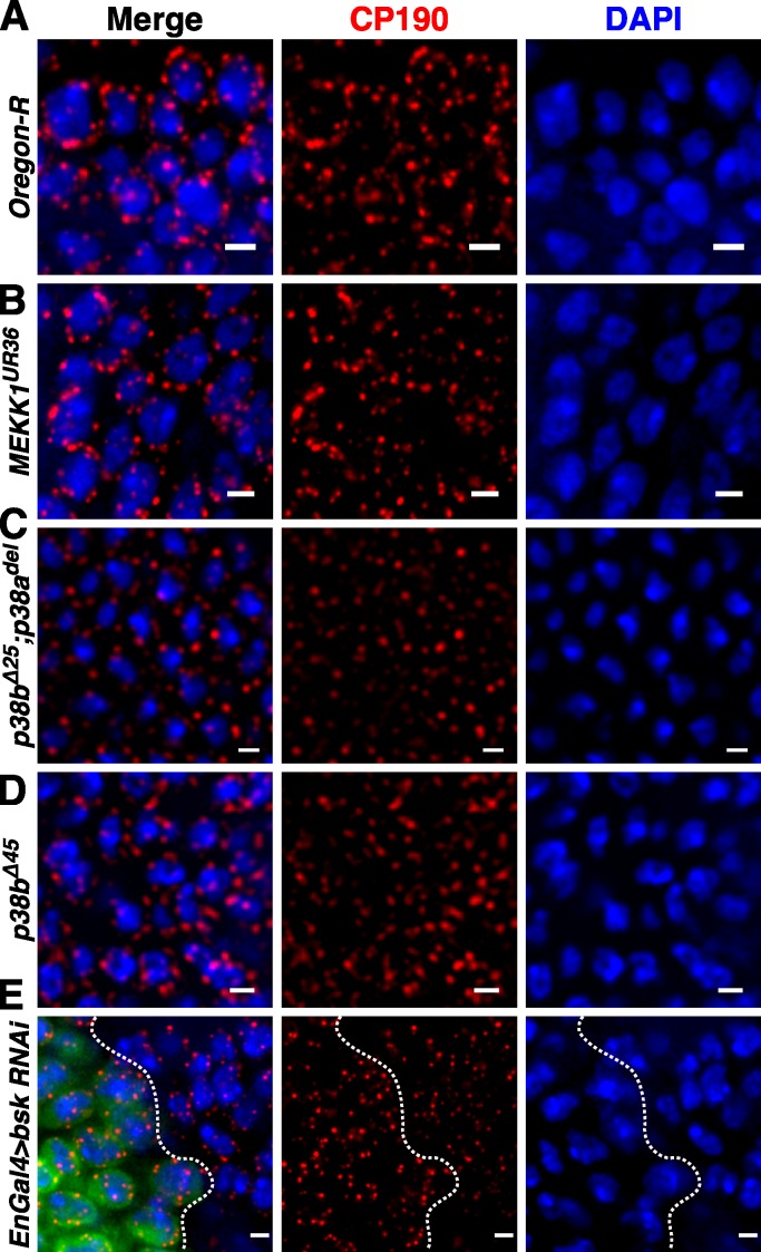
Chromatin insulators assist in the formation of higher-order chromatin structures by mediating long-range contacts between distant genomic sites. It has been suggested that insulators accomplish this task by forming dense nuclear foci termed insulator bodies that result from the coalescence of multiple protein-bound insulators. However, these structures remain poorly understood, particularly the mechanisms triggering body formation and their role in nuclear function. In this paper, we show that insulator proteins undergo a dramatic and dynamic spatial reorganization into insulator bodies during osmostress and cell death in a high osmolarity glycerol-p38 mitogen-activated protein kinase-independent manner, leading to a large reduction in DNA-bound insulator proteins that rapidly repopulate chromatin as the bodies disassemble upon return to isotonicity. These bodies occupy distinct nuclear territories and contain a defined structural arrangement of insulator proteins. Our findings suggest insulator bodies are novel nuclear stress foci that can be used as a proxy to monitor the chromatin-bound state of insulator proteins and provide new insights into the effects of osmostress on nuclear and genome organization.


Figures

Figure 1.
Figure 1.
Insulator bodies form in response to osmostress. (A and B) S2 cells stained with CP190 and Mod(mdg4) under normal cellular conditions (A) or after treatment with 250 mM NaCl (B). (C and D) Wing discs from third instar larvae stained for CP190 and Mod(mdg4) under normal cellular conditions (C) or after treatment with 250 mM NaCl (D). (E and F) Orthogonal projections along the indicated axes (yellow dashed lines) in an unstressed (E) and stressed (F) S2 cell. The red box outlines the x-y plane image. Note that A–D are maximum projections of 1-µm z slices, whereas E and F are a single z slice (x-y plane only). Bars, 2 µm.
Figure 2.
Figure 2.
Osmostress does not alter the nuclear distribution of other chromatin proteins. (A and B) S2 cells treated with or without 250 mM NaCl and stained for CP190 and Polycomb (A) or HP1 (B). Bars, 2 µm.
Figure 3.
Figure 3.
Insulator bodies have a defined structural organization. (A and B) S2 cells treated with or without 250 mM NaCl and stained for CP190 and Su(Hw) (A), dCTCF::mCherry and Su(Hw)::EGFP (B) and BEAF-32 and Su(Hw) after 60-min incubation in PBS. (C) BEAF-32 forms large donut structures around the spherical structures (arrowhead and insets). Bars: (A–C, main images) 2 µm; (C, insets) 0.5 µm.
Figure 4.
Figure 4.
Insulator bodies localize to DAPI-less regions and associate transiently with the nuclear lamina. S2 cells treated with or without 250 mM NaCl and stained for CP190 and lamin. (A) Bodies localized to the interior form in DAPI-less lacunas (white arrowheads). Intensity correlation analysis (boxed regions and insets) reveals regions of high overlap (gold) between CP190 bodies in the nuclear periphery and lamin. (B) Serial 1-µm z slices through a S2 nucleus stressed with 250 mM NaCl stained for CP190 and nuclear pore complex components (NUPs). (C) Nuclear halos generated from 250 mM NaCl-stressed S2 cells showing a highly extracted nucleus with no CP190 signal (yellow asterisks) and a less efficiently extracted nucleus (white asterisks) showing remnants of CP190 bodies colocalized with lamin. Bars: (A, main images) 2 µm; (A, insets) 0.5 µm; (B) 1 µm; (C) 4 µm.
Figure 5.
Figure 5.
Buffer choice and tissue dissection conditions can account for insulator body formation. (A and B) Wing discs from third instar larvae dissected in SFX media, PBS, or Drosophila Ringer’s solution and fixed in <5 min (A) or after a 30-min incubation in nonhumidified conditions (B), stained for CP190 and Mod(mdg4). (C) S2 cells show a similar response after a 30-min incubation in PBS or Ringer’s solution (media controls were kept humidified to prevent evaporation). Bars, 2 µm.
Figure 6.
Figure 6.
Insulator body formation and disassembly occurs rapidly, and bodies are highly dynamic. (A and B) Frames taken at 2-min intervals after gradual 250 mM NaCl media addition at time 0 min in S2 cells expressing Su(Hw)::EGFP and BEAF-32::mCherry. Bodies form in a matter of minutes from diffuse speckles (A) and can undergo rounds of fusion (bodies 1 and 2) to produce larger structures (body 3*; B). Boxed regions are enlarged in insets at the bottom. (C) The dynamic movement of body 1 starting with its formation at 4 min until its fusion with body 2 at 20 min (blue line) and the movement of the fused body (white line) until the final frame was acquired (36 min). (D) A polytene nucleus from a third instar salivary gland expressing Su(Hw)::EGFP subjected to 250 mM NaCl osmostress (10 min) followed by recovery in isotonic media (23 min). Blue numbers denote media treatment time points, and green numbers indicate stress treatment time points. Arrowheads (9 and 25 min) mark bands of Su(Hw). Bars: (A, B [top], and D) 3 µm; (B [bottom] and C) 2 µm. Also see Videos 1, 2, 3, and 4.
Figure 7.
Figure 7.
Insulator body formation correlates with a reduction in chromatin-bound Su(Hw) that is rapidly restored upon return to isotonicity. (A) A media-treated salivary gland polytene nucleus labeled with CP190 showing the expected band pattern (insets, arrowheads). (B) A polytene nucleus stressed with 250 mM NaCl labeled with CP190 shows bodies in the nuclear periphery and interchromosomal spaces lacking DAPI (insets). (A and B) Boxed regions are enlarged in insets on the right. (C–E) ChIP of Su(Hw) at gypsy (C), 3L:12247800 (D), and homie insulators (E) in media, stressed, and recovery S2 cells. Asterisks mark reductions significantly different from media controls (Student’s paired t test, P = 0.05; error bars represent SEMs). Bars: (A and B, main images) 3 µm; (A and B, insets) 0.5 µm.
Figure 8.
Figure 8.
CP190 and Mod(mdg4) enter bodies independently of one another, but Su(Hw) requires Mod(mdge4) in larval tissue. (A) Wing discs from CP190-RNAi larvae stressed with 250 mM NaCl and labeled with CP190 (left) or Mod(mdg4) (middle). Unstressed controls labeled with Mod(mdg4) are shown on the right. GFP marks Dcr-2+ knockdown cells, and the dashed lines demarcate the anterior–posterior axis of the wing disc. (B) Wing discs from two trans-heterozygous CP190 mutant larvae, CP190H31-2/CP190P11 (left) and CP1904-1/CP190P11 (middle) and a balanced control containing full-length CP190 (right) stressed with 250 mM NaCl and stained with CP190 and Mod(mdg4). Note that our CP190 antibody recognizes the CP1904-1 isoform but not the CP190H31-2 isoform. Domains of CP190 (BTB, Asp rich [Asp], microtubule binding [Cen], Zinc finger [Znf], and Glu rich [Glu]) present in each truncated allele are indicated by the colored lines. (C) Wing discs from null modu1 homozygotes (left) and balanced heterozygotes (middle) stressed with 250 mM NaCl and stained with Su(Hw) and CP190. (right) Mod staining verifies absence of protein in the modu1 mutant. Bars, 2 µm.
Figure 9.
Figure 9.
Insulator body formation is independent of the HOG–MAPK osmostress pathway. (A–E) Wing discs from Oregon-R (A), dMEKK1UR36/dMEKKUR36 (B), p38bΔ25;p38adel/p38bΔ25;p38adel (C), p38bΔ45/p38bΔ45 (D), and bsk (JNK)-RNAi (GFP marks Dcr-2+ knockdown cells, and the dashed lines demarcate the anterior–posterior axis of the wing disc; E) stressed with 250 mM NaCl and stained with CP190. Bars, 2 µm.
Figure 10.
Figure 10.
A model for insulator body formation during osmostress and cell death. NUP, nuclear pore complex.

Similar articles

Cited by

References

    1. Alkema M.J., Bronk M., Verhoeven E., Otte A., van ’t Veer L.J., Berns A., van Lohuizen M. 1997. Identification of Bmi1-interacting proteins as constituents of a multimeric mammalian polycomb complex. Genes Dev. 11:226–240 10.1101/gad.11.2.226 - DOI - PubMed
    1. Bantignies F., Roure V., Comet I., Leblanc B., Schuettengruber B., Bonnet J., Tixier V., Mas A., Cavalli G. 2011. Polycomb-dependent regulatory contacts between distant Hox loci in Drosophila. Cell. 144:214–226 10.1016/j.cell.2010.12.026 - DOI - PubMed
    1. Biamonti G., Vourc’h C. 2010. Nuclear stress bodies. Cold Spring Harb. Perspect. Biol. 2:a000695 10.1101/cshperspect.a000695 - DOI - PMC - PubMed
    1. Burg M.B., Ferraris J.D., Dmitrieva N.I. 2007. Cellular response to hyperosmotic stresses. Physiol. Rev. 87:1441–1474 10.1152/physrev.00056.2006 - DOI - PubMed
    1. Bushey A.M., Ramos E., Corces V.G. 2009. Three subclasses of a Drosophila insulator show distinct and cell type-specific genomic distributions. Genes Dev. 23:1338–1350 10.1101/gad.1798209 - DOI - PMC - PubMed

Publication types

MeSH terms

LinkOut - more resources