. 2013 Aug 15;191(4):1529-35.
doi: 10.4049/jimmunol.1300284. Epub 2013 Jul 22.

Allergens as immunomodulatory proteins: the cat dander protein Fel d 1 enhances TLR activation by lipid ligands

Affiliations

Allergens as immunomodulatory proteins: the cat dander protein Fel d 1 enhances TLR activation by lipid ligands

Jurgen Herre et al. J Immunol. .

Abstract

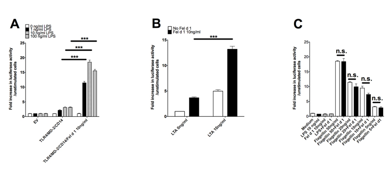
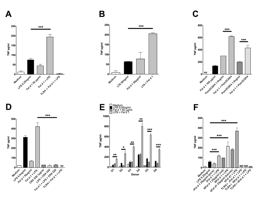
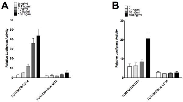
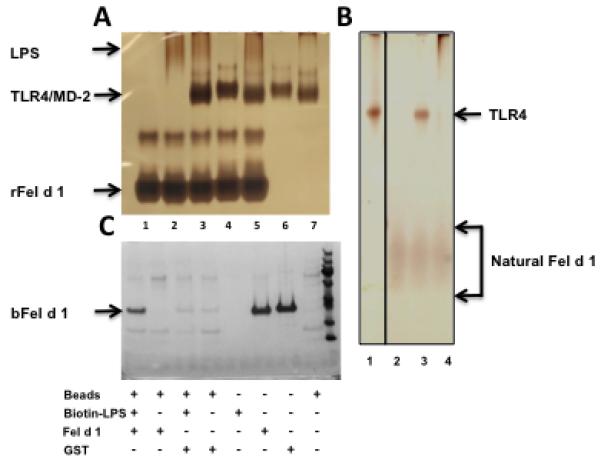
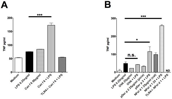
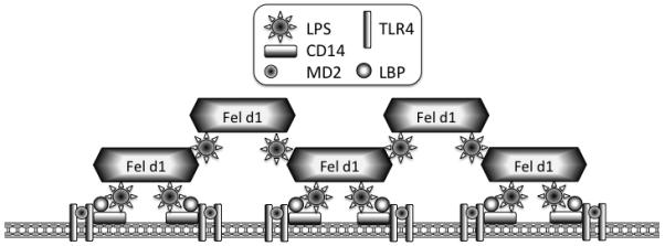
Allergic responses can be triggered by structurally diverse allergens. Most allergens are proteins, yet extensive research has not revealed how they initiate the allergic response and why the myriad of other inhaled proteins do not. Among these allergens, the cat secretoglobulin protein Fel d 1 is a major allergen and is responsible for severe allergic responses. In this study, we show that similar to the mite dust allergen Der p 2, Fel d 1 substantially enhances signaling through the innate receptors TLR4 and TLR2. In contrast to Der p 2, however, Fel d 1 does not act by mimicking the TLR4 coreceptor MD2 and is not able to bind stably to the TLR4/MD2 complex in vitro. Fel d 1 does, however, bind to the TLR4 agonist LPS, suggesting that a lipid transfer mechanism may be involved in the Fel d 1 enhancement of TLR signaling. We also show that the dog allergen Can f 6, a member of a distinct class of lipocalin allergens, has very similar properties to Fel d 1. We propose that Fel d 1 and Can f 6 belong to a group of allergen immunomodulatory proteins that enhance innate immune signaling and promote airway hypersensitivity reactions in diseases such as asthma.


Figures

Figure 1
Figure 1. Fel d 1 enhances signalling by ligands of TLR2 and 4 but not TLR5
(A) HEK293 cells transiently transfected to express TLR4, CD14, MD2, an NFkB-luciferase reporter (pNFkB-luc) and a constitutively active luciferase reporter control (phRG-TK) were treated for 6 hours with LPS (1-100 ng/ml) in the presence or absence of Fel d 1 (10 ng/ml). Cells were lysed and the NFkB luciferase reporter was read on a FLUOstar Omega (BMG Labtech) luminometer. Data are normalized against the unstimulated control. The graph is one representative experiment from 4 separate experiments. (B) HEK293 cells transiently transfected with TLR2, CD14 pNFkB-luc and phRG-TK were stimulated with lipotechoic acid (LTA) at 10ng/ml in the presence, and absence, of Fel d 1 (10 ng/ml). Data are normalized against the unstimulated control. Data are shown from one representative experiment from 3 separate experiments. (C) HEK293 cells were transiently transfected with TLR5, NFkB-luc and phRG-TK) then stimulated with flagellin (5-50 ng/ml) in the presence or absence of Fel d 1 (10 ng/ml). Data normalized against unstimulated control are shown as one representative experiment from 5 separate experiments. Data represent mean and s.e.m. of a representative experiment from 4-5 repeat experiments. *P<0.05; **P<0.005;***P<0.001; n.s., not significant.
Figure 2
Figure 2. Fel d 1 enhances expression of the pro-inflammatory cytokine TNF-alpha in primary cells of the innate immune system
(A) Bone marrow derived macrophages (BMDM) from wild type and TLR4−/− C57BL6 mice were treated for 24h with LPS at 0.5ng/ml, Fel d 1 at 50μg/ml or both, as indicated for 24 hours. TNF-alpha levels were measured by an ELISA (R&D). Data from 5 separate experiments was pooled and expressed as mean±sem. (B) Wild type BMDMs were treated with LTA at 50ng/ml with or without Fel d 1 at 50μg/ml for 24 hours. TNF-alpha levels were measured by an ELISA (R&D). Data from 3 separate experiments were pooled and expressed as mean±sem. (C) Wild-type BMDMs treated for 24h with di- and tri acylated lipid activators of TLR1/2 and TLR2/6 (Pam3CSK4 1.0ng/ml and Pam2CSK4 1.0ng/ml) with or without Fel d 1 at 100μg/ml. TNF-alpha levels were measured by an ELISA (R&D). Data from 2 separate experiments were pooled and expressed as mean±sem. (D) Wild type and TLR4−/− BMDMs were pre-treated with the TLR4 small molecule inhibitor CRX-526 for 60 minutes and then treated for 24h with LPS 0.5ng/ml with or without Fel d 1 at 50μg/ml. CRX-526 was maintained throughout the stimulation period. TNF-alpha levels were measured by an ELISA (R&D). Data from 2 separate experiments were pooled and expressed as mean±sem. (E) Peripheral Blood Mononuclear Cells (PBMC) derived from healthy donors as part of an ethically approved research programme were treated for 24h with LPS 0.05ng/ml with or without Fel d 1 at 50μg/ml. Donors are shown separately to demonstrate inter individual variation. TNF-alpha levels were measured by an ELISA (R&D). Data are shown from 3 healthy donors. (F) BMDM from wild type and TLR4−/− mice treated with LPS 0.25ng/ml and six alternative sources of Fel d 1 (recombinant protein expressed in the yeast Pichia pastoris (rFel d 1), natural protein from the cat depleted of LPS (NFel d 1) and Fel d 1 made in baculovirus (bFel d 1)) each at 25μg/ml. TNF-alpha levels were measured by an ELISA (R&D). Data from 3 separate experiments were pooled and expressed as mean±sem. *P<0.05; **P<0.005;***P<0.001; n.s., not significant
Figure 3
Figure 3. Signal enhancement by Fel d 1 requires the TLR4 co-receptor MD2 and CD14
(A) HEK293 cells were transfected with components of the TLR4 signalling complex: either TLR4, MD2 and CD14; or TLR4 and CD14, an NFkB-luciferase reporter (pNFkB-luc) and a constitutively active luciferase reporter control (phRG-TK) and then stimulated with Fel d 1 (0.1-100ng/ml). The graph is one representative experiment from 3 separate experiments. (B) HEK293 cells were transfected with components of the TLR4 signalling complex: either TLR4, MD2 and CD14; or TLR4 and MD2 an NFkB-luciferase reporter (pNFkB-luc) and a constitutively active luciferase reporter control (phRG-TK) and then stimulated with Fel d 1 (0.1-100ng/ml). The graph is one representative experiment from 3 separate experiments.
Figure 4
Figure 4. Fel d 1 interacts with LPS, but not with the TLR4/MD2 complex
(A) Recombinant Fel d 1 does not interact with TLR4/MD2. Silver-stained gel of recombinant Fel d 1 (rFel d 1) that has been incubated with LPS and components of a co-purified TLR4/MD2 complex. 1 μg of each component was added in the order listed and incubated at room temperature for 30 min: 1 = rFel d 1alone; 2 = rFel d 1 + LPS; 3 = rFel d 1 + LPS + TLR4/MD2; 4 =rFel d 1 + TLR4/MD2; 5 = rFel d 1 + TLR4/MD2 + LPS; 6 = TLR4/MD2; 7 = TLR4/MD2 + LPS. The position of migration for each component is shown. rFel d 1 migrates as two bands consistent with the formation of a dimer and tetramer. (B) Natural Fel d 1 does not form a complex with TLR4 either in the presence or absence of LPS. 1 μg of each of the listed components were incubated together at room temperature for 30 minutes: 1 = TLR4 alone; 2 = natural Fel d 1 alone; 3 = natural Fel d 1 + TLR4; 4 = natural Fel d 1 + LPS + TLR4. No shift in the band for natural Fel d 1 was observed. (C) Fel d 1 interacts with LPS. Recombinant Fel d 1 from baculovirus (bFel d 1) was incubated with biotin-LPS and the complex pulled down using streptavidin-conjugated beads (lane 1). The position of bFel d 1 is marked. Lanes 2, 3 and 4 control for non-specific binding of bFel d 1 to the streptavidin-coated beads; non-specific binding of proteins to biotin-LPS; and non-specific binding of GST to the streptavidin-coated beads. Lanes 5 to 8 indicate assay inputs.
Figure 5
Figure 5. Allergens from three different structural classes sensitize signalling by TLR4 and TLR2
(A) Wild-type and TLR4−/− BMDM treated with LPS at 0.25ng/ml in the presence, or absence, of the dog lipocalin allergen Can f 6 at 50μg/ml for 24 hours. TNF-alpha levels were measured by an ELISA (R&D). Data from 3 separate experiments were pooled and expressed as mean±sem. (B) Wild-type and TLR4−/− BMDMs treated with LPS (0.25ng/ml), the house dust mite allergen Der p 2 (25μg/ml), the model allergen protein ovalbumin (OVA, 20μg/ml) or cat derived Fel d 1 (NFel d 1, 25μg/ml) either singly or in combination for 24h. TNF-alpha levels were measured by an ELISA (R&D). Data from 3 separate experiments were pooled and expressed as mean±sem. *P<0.05; **P<0.005;***P<0.001; n.s., not significant.
Figure 6
Figure 6. Model of LPS sensitization by dander proteins
Animal dander proteins loaded with environmentally derived lipid pathogen-associated molecular patterns (PAMPs) associate with cell membrane, facilitating lipid presentation or transfer to receptor complexes. Dander proteins, in the presence of low LPS concentrations, could cluster together with LPS to form larger complexes that then promote greater clustering of TLR4 bearing lipid rafts, leading to increased receptor activation. At higher LPS exposure levels (such as would not be seen from environmental sources), this effect is not seen because maximal receptor activation has already been achieved.

References

    1. Minnicozzi M, Sawyer RT, Fenton MJ. Innate immunity in allergic disease. Immunological reviews. 2011;242:106–127. - PubMed
    1. Spitzauer S. Allergy to mammalian proteins: at the borderline between foreign and self? International archives of allergy and immunology. 1999;120:259–269. - PubMed
    1. Chiou Y-L, Lin C-Y. Der p2 activates airway smooth muscle cells in a TLR2/MyD88-dependent manner to induce an inflammatory response. Journal of cellular physiology. 2009;220:311–318. - PubMed
    1. Trompette A, Divanovic S, Visintin A, Blanchard C, Hegde RS, Madan R, Thorne PS, Wills-Karp M, Gioannini TL, Weiss JP, Karp CL. Allergenicity resulting from functional mimicry of a Toll-like receptor complex protein. Nature. 2009;457:585–588. - PMC - PubMed
    1. Wilson RH, Maruoka S, Whitehead GS, Foley JF, Flake GP, Sever ML, Zeldin DC, Kraft M, Garantziotis S, Nakano H, Cook DN. The Toll-like receptor 5 ligand flagellin promotes asthma by priming allergic responses to indoor allergens. Nature medicine. 2012;18:1705–1710. - PMC - PubMed

Publication types

MeSH terms