

Conjunctival HLA-DR and CD8 expression detected by impression cytology in ocular graft versus host disease
- PMID: 23878500
- PMCID: PMC3716410
Conjunctival HLA-DR and CD8 expression detected by impression cytology in ocular graft versus host disease
Abstract
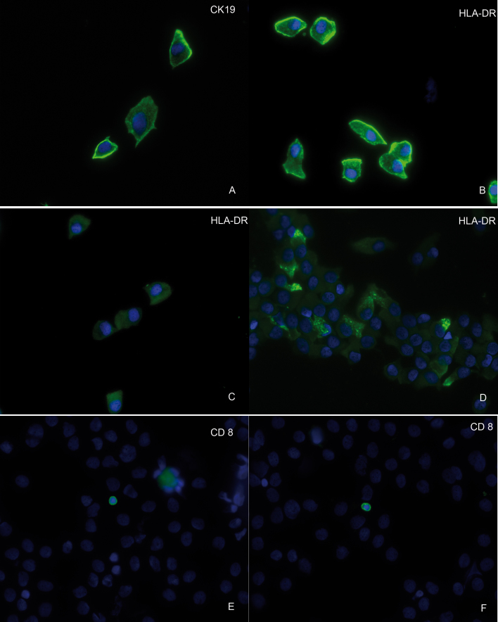
Purpose: To assess the expression of human leucocyte antigen (HLA)-DR in epithelial cells and cluster of differentiation (CD8)-positive lymphocytes as possible markers of chronic ocular graft versus host disease (cGvHD) after hematological stem cell transplantation (HSCT).
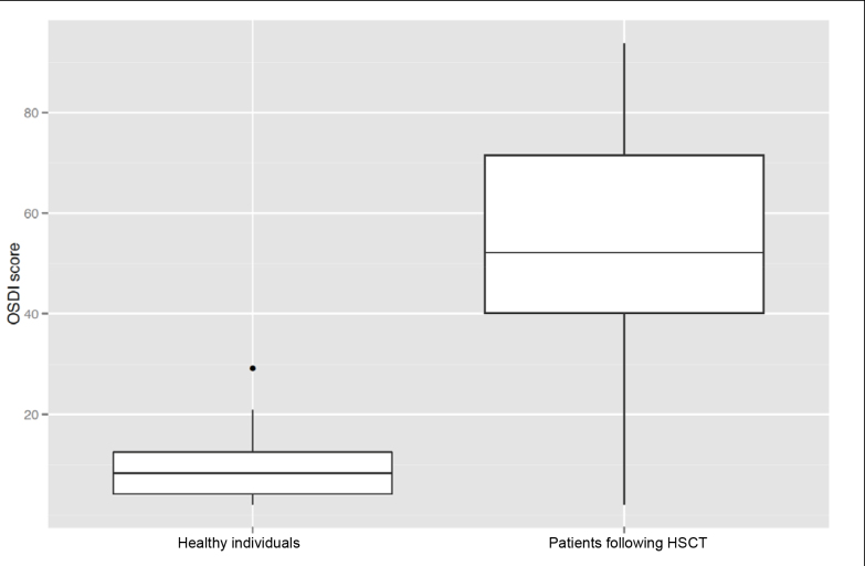
Methods: Twenty-seven consecutive patients with dry-eye symptoms following HSCT (24 [89%] with peripheral blood stem cell transplantation and 3 [11%] with bone marrow transplants; 17 [63%] familiar allogenic grafts) and 19 age-matched controls were included. Conjunctival impression cytology specimens were stained for HLA-DR, cytokeratin 19, and CD8. Oxford grading scale, blinking frequency, Schirmer test, tear film break-up time (TBUT), and Ocular Surface Disease Index (OSDI) were also recorded. Wilcoxon nonparametric testing was used to compare controls and HSCT recipients and to assess HSCT recipient subgroups with and without clinical cGVHD.
Results: Eighteen patients showed clinical signs of ocular cGVHD. TBUT and Schirmer test scores were significantly lower in patients, while Oxford grades and OSDI were significantly higher than in controls. Epithelial HLA-DR expression was generally higher in HSCT recipients than in controls, but it did not correlate with ocular cGVHD status. CD8-positive lymphocytes were identified in five patients with ocular cGvHD and one control.
Conclusions: A strong HLA-DR expression as detected by impression cytology appears to indicate a general HSCT response and fails to predict ocular cGVHD. However, the detection of CD8-positive lymphocytes using impression cytology was frequently associated with ocular cGvHD. Our data warrant further evaluation of CD8 expression in impression cytology, along with comparison to conjunctival biopsies and brush cytology, as impression cytology may offer a less invasive strategy for assessing cGVHD status.
Figures


Similar articles
-
A study of conjunctival impression cytology in patients undergoing allogeneic hematopoietic stem cell transplantation and its relationship with Ocular Graft versus Host Disease.Rom J Ophthalmol. 2025 Jan-Mar;69(1):68-73. doi: 10.22336/rjo.2025.12. Rom J Ophthalmol. 2025. PMID: 40330961 Free PMC article.
-
Baseline profiles of ocular surface and tear dynamics after allogeneic hematopoietic stem cell transplantation in patients with or without chronic GVHD-related dry eye.Bone Marrow Transplant. 2010 Jun;45(6):1077-83. doi: 10.1038/bmt.2009.312. Epub 2009 Nov 9. Bone Marrow Transplant. 2010. PMID: 19898506 Clinical Trial.
-
Tear function and lipid layer alterations in dry eye patients with chronic graft-vs-host disease.Eye (Lond). 2009 Jan;23(1):202-8. doi: 10.1038/eye.2008.340. Epub 2008 Nov 14. Eye (Lond). 2009. PMID: 19011604
-
Ocular graft-versus-host disease: Risk factors of ocular graft-versus-host disease after allogeneic haematopoietic stem cell transplantation in Denmark.Acta Ophthalmol. 2025 Apr;103 Suppl 286(Suppl 286):3-19. doi: 10.1111/aos.17452. Acta Ophthalmol. 2025. PMID: 40211651 Free PMC article. Review.
-
Review of Genetic Variation as a Predictive Biomarker for Chronic Graft-Versus-Host-Disease After Allogeneic Stem Cell Transplantation.Front Immunol. 2020 Oct 19;11:575492. doi: 10.3389/fimmu.2020.575492. eCollection 2020. Front Immunol. 2020. PMID: 33193367 Free PMC article.
Cited by
-
Aberrant HLA-DR expression in the conjunctival epithelium after autologous serum treatment in patients with graft-versus-host disease or Sjögren's syndrome.PLoS One. 2020 Apr 21;15(4):e0231473. doi: 10.1371/journal.pone.0231473. eCollection 2020. PLoS One. 2020. PMID: 32315325 Free PMC article.
-
A Clinical Trial Comparing the Safety and Efficacy of Topical Tacrolimus versus Methylprednisolone in Ocular Graft-versus-Host Disease.Ophthalmology. 2016 Jul;123(7):1449-57. doi: 10.1016/j.ophtha.2016.02.044. Epub 2016 Apr 13. Ophthalmology. 2016. PMID: 27086024 Free PMC article. Clinical Trial.
-
Recent advances in ocular graft-versus-host disease.Front Immunol. 2023 Jan 25;14:1092108. doi: 10.3389/fimmu.2023.1092108. eCollection 2023. Front Immunol. 2023. PMID: 36761771 Free PMC article. Review.
-
Comparison among different diagnostic criteria for chronic ocular graft-versus-host disease applied with and without pre-transplant ophthalmological examination.Eye (Lond). 2019 Jan;33(1):154-160. doi: 10.1038/s41433-018-0210-4. Epub 2018 Sep 7. Eye (Lond). 2019. PMID: 30194377 Free PMC article.
-
Review of Biomarkers in Ocular Matrices: Challenges and Opportunities.Pharm Res. 2019 Jan 23;36(3):40. doi: 10.1007/s11095-019-2569-8. Pharm Res. 2019. PMID: 30673862 Free PMC article. Review.
References
-
- Holler E. Risk assessment in haematopoietic stem cell transplantation: GvHD prevention and treatment. Best Pract Res Clin Haematol. 2007;20:281–94. - PubMed
-
- Saito T, Shinagawa K, Takenaka K, Matsuo K, Yoshino T, Kiura K, Niiya K, Harada M. Ocular manifestation of acute graft-versus-host disease after allogeneic peripheral blood stem cell transplantation. Int J Hematol. 2002;75:332–4. - PubMed
-
- Riemens A, te Boome L, Imhof S, Kuball J, Rothova A. Current insights into ocular graft-versus-host disease. Curr Opin Ophthalmol. 2010;21:485–94. - PubMed
-
- Wang Y, Ogawa Y, Dogru M, Tatematsu Y, Uchino M, Kamoi M, Okada N, Okamoto S, Tsubota K. Baseline profiles of ocular surface and tear dynamics after allogeneic hematopoietic stem cell transplantation in patients with or without chronic GVHD-related dry eye. Bone Marrow Transplant. 2010;45:1077–83. - PubMed
-
- Rojas B, Cuhna R, Zafirakis P, Ramirez JM, Lizan-garciia M, Zhao T, Foster CS. Cell populations and adhesion molecules expression in conjunctiva before and after bone marrow transplantation. Exp Eye Res. 2005;81:313–25. - PubMed
MeSH terms
Substances
LinkOut - more resources
Full Text Sources
Research Materials