. 2013 Jul 19:19:1526-37.
Print 2013.

Keratoconus corneal architecture after riboflavin/ultraviolet A cross-linking: ultrastructural studies

Affiliations

Keratoconus corneal architecture after riboflavin/ultraviolet A cross-linking: ultrastructural studies

Saeed Akhtar et al. Mol Vis. .

Abstract

Purpose: Study to investigate the effects of collagen cross-linking on the ultrastructural organization of the corneal stroma in the human keratoconus cornea (KC).

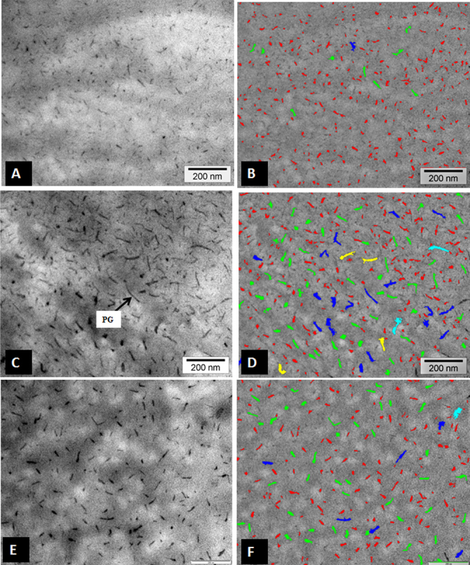
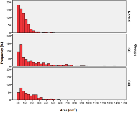
Methods: Three normal, three keratoconus (KC1, KC2, KC3), and three cross-linked keratoconus (CXL1, CXL2, CXL3) corneas were analyzed. The KC corneas were treated with a riboflavin-ultraviolet A (UVA) treatment (CXL) method described by Wollensak et al. Penetrating keratoplasty (PKP) was performed 6 months after treatment. All samples were processed for electron microscopy.

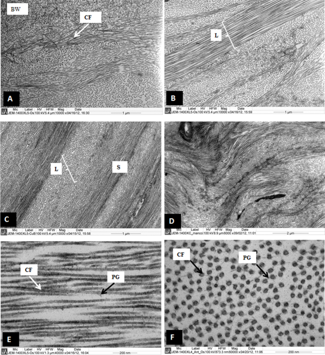
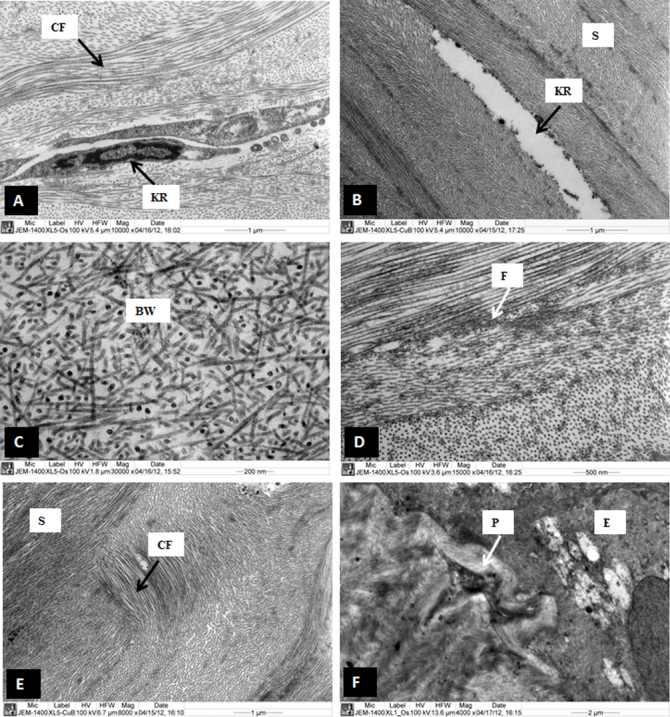
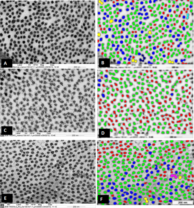
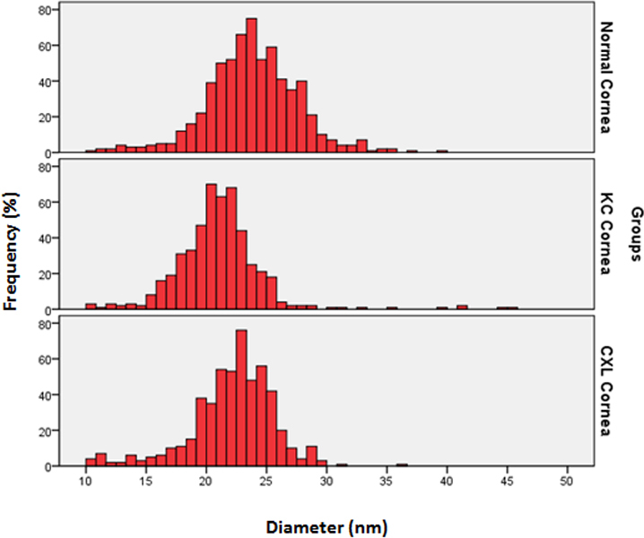
Results: The riboflavin-UVA-treated CXL corneal stroma showed interlacing lamellae in the anterior stroma followed by well-organized parallel running lamellae. The lamellae contained uniformly distributed collagen fibrils (CFs) decorated with normal proteoglycans (PGs). The CF diameter and interfibrillar spacing in the CXL cornea were significantly increased compared to those in the KC cornea. The PG area in the CXL corneas were significantly smaller than the PGs in the KC cornea. The epithelium and Bowman's layer were also normal. On rare occasions, a thick basement membrane and collagenous pannus were also observed.

Conclusions: Corneal cross-linking leads to modifications of the cornea stroma. The KC corneal structure showed a modification in the CF diameter, interfibrillar spacing, and PG area. This resulted in a more uniform distribution of collagen fibrils, a key feature for corneal transparency.


Figures

Figure 1
Figure 1
Electron micrograph of cross-linked sample 1 and keratoconus corneas. A: Collagen fibrils are emerging from the posterior part of the Bowman’s layer. B: Interlacing lamellae are present in the anterior stroma. C: Parallel running collagen fibril lamellae are present in the posterior part of the anterior stroma. D: Part of the keratoconus (KC) corneas are showing undulating lamellae. E: Longitudinally running collagen fibrils are decorated with proteoglycan (PGs), in the posterior part of anterior stroma. F: Cross section of the collagen fibrils are decorated with PGs in the anterior part of the stroma. BW= Bowman’s layer, CF=Collagen fibrils, L=Lamella, PG=Proteoglycans, S=Stroma.
Figure 2
Figure 2
Electron micrograph of sample cross-linked (CXL) samples 1, 2, and 3. A: A normal keratocyte with large nucleus is present in the anterior part of the stroma of CXL1. B: An electron lucent part of keratocyte is present in the anterior part of the stroma of CXL3. C: A part of Bowman’s layer which appeared normal in CXL1. D: Patches of microfilaments are present in the anterior stroma of CXL1. E: Randomly running collagen fibrils are present in the anterior stroma in the anterior stroma of CXL1. F: A collagenous pannus at sub-epithelial region is pushing basal epithelial cell in CXL2. E=Epithelium, BW=Bowman’s layer, CF=Collagen fibrils, F=Micro-filaments, KR=Keratocyte, p=Pannus, S=Stroma.
Figure 3
Figure 3
Electron micrograph and digital images of the collagen fibrils of normal, keratoconus (KC), and cross-linked (CXL) corneas. A: Electron micrograph of collagen fibrils which are present in the normal human corneas. B: Digital image obtained after processing the image shown in A. C: Electron micrograph of the collagen fibrils which are present in the KC corneas. D: Digital image is obtained after processing the image shown in C. E: Electron micrograph of collagen fibrils which are present in the CXL corneas. F: Digital image obtained after processing the image shown in E. The images were displayed by using color coding to demonstrate the distribution of the variable diameters of collagen fibrils. Collagen fibril color coding: Red=10–15 nm, Green=15–20 nm, Blue=20–25 nm, Yellow=24–30 nm, Aqua=30–35 nm, Pink=35–40 nm, Brown=40–45 nm.
Figure 4
Figure 4
Distribution of collagen fibril diameter (nm) in normal corneas, keratoconus (KC) corneas, and cross-linked (CXL) corneas. The data were derived from three corneas and six images of each group.
Figure 5
Figure 5
Electron micrograph and digital images of proteoglycans (PG) of normal, keratoconus (KC), and cross-linked (CXL) corneas. A: Electron micrograph of PGs which are present of normal corneas. B: Digital image is obtained after processing image shown in A, showing variable area distribution of PGs. C: Electron micrograph which are present in PGs of KC corneas. D: Digital image is obtained after processing image shown in C, showing variable area distribution of PGs. E: Electron micrograph of PGs of CXL corneas. F: Digital image obtained after processing image shown in E, showing variable area distribution of PGs. PG=Proteoglycan Proteoglycans color coding: Red=50–350 nm2, Green=350–650 nm2, Blue=650–950 nm2, Yellow=950–1250 nm2, Aqua=1250–1550 nm2, Pink=1550–1850 nm2, Brown=1850–2150 nm2.
Figure 6
Figure 6
Distribution of proteoglycan area (nm2) in normal, keratoconus, and cross-linked corneas. The data were derived from three corneas and six images of each group.

Similar articles

Cited by

References

    1. Rabinowitz YS. Keratoconus. Surv Ophthalmol. 1998;42:297–319. - PubMed
    1. Tuft SJ, Moodaley LC, Gregory WM, Davison CR, Buckley RJ. Prognostic factors for the progression of keratoconus. Ophthalmology. 1994;101:439–47. - PubMed
    1. Kennedy RH, Bourne WM, Dyer JA. A 48-year clinical and epidemiologic study of keratoconus. Am J Ophthalmol. 1986;101:267–73. - PubMed
    1. Wollensak G, Spoerl E, Seiler T. Riboflavin/ultraviolet-a-induced collagen crosslinking for the treatment of keratoconus. Am J Ophthalmol. 2003;135:620–7. - PubMed
    1. Caporossi A, Mazzotta C, Baiocchi S, Caporossi T. Long-term results of riboflavin ultraviolet a corneal collagen cross-linking for keratoconus in Italy: the Siena eye cross study. Am J Ophthalmol. 2010;149:585–93. - PubMed

Publication types

LinkOut - more resources