

Progressive hemifacial atrophy
- PMID: 23878573
- PMCID: PMC3714811
- DOI: 10.4103/1735-3327.111810
Progressive hemifacial atrophy
Abstract
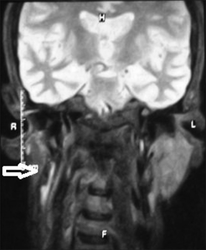
Progressive hemifacial atrophy, also known as Parry-Romberg Syndrome, is an uncommon degenerative and poorly understood condition. It is characterized by a slow and progressive but self-limited atrophy affecting one side of the face. The incidence and the cause of this alteration are unknown. A cerebral disturbance of fat metabolism has been proposed as a primary cause. Possible factors that are involved in the pathogenesis include trauma, viral infections, heredity, endocrine disturbances and auto-immunity. The most common complications that appear in association to this disorder are: trigeminal neuralgia, facial paresthesia, severe headache and epilepsy. Characteristically, the atrophy progresses slowly for several years and, it becomes stable. The objective of this work is, through the presentation of a clinical case, to accomplish a literature review concerning general characteristics, etiology, physiopathology and treatment of progressive hemifacial atrophy.
Keywords: Parry-Romberg Syndrome; Progressive hemifacial atrophy; Romberg's disease.
Conflict of interest statement
Figures


References
-
- Thiago Pastor da Silva Pinheiro, Camila Camarinha da Silva, Carolina Souza Limeira da Silveira, Patrícia Cristina Ereno Botelho, Maria das Graças Rodrigues Pinheiro, Joao de Jesus Viana Pinheiro. Progressive hemifacial atrophy – Case report. Med Oral Patol Oral Cir Bucal. 2006;11:E112–4. - PubMed
-
- Pensler JM, Murphy GF, Muliken JB. Clinical and ultra-structural studies of Romberg's hemifacial atrophy. Plast Reconstr Surg. 1990;85:669–76. - PubMed
-
- Mazzeo N, Fisher JG, Mayer MH, Mathieu GP, Mcade FG. Progressive hemifacial atrophy (Parry Romberg Syndrome) Oral Surg Oral Med Oral Pathol Oral Radiol Endod. 1995;79:30–5. - PubMed
-
- Neville BW, Damm DD, Allen CN, Bouqout JE. Facial surgical: Pathology Oral Maxilofacial. In: Koogan G, editor. 5th ed. Vol. 1. Rio de Janeiro: Nike and Lidman; 1998. pp. 35–42.
Publication types
LinkOut - more resources
Full Text Sources