

Tpeak -tend interval in 12-lead electrocardiogram of healthy children and adolescents tpeak -tend interval in childhood
- PMID: 23879274
- PMCID: PMC6932219
- DOI: 10.1111/anec.12035
Tpeak -tend interval in 12-lead electrocardiogram of healthy children and adolescents tpeak -tend interval in childhood
Abstract
Background: Tpeak (Tp) to the Tend (Te) interval is an index of transmural dispersion of repolarization. Prolongation of this interval predisposes to life-threatening ventricular arrhythmias in long QT syndrome, polymorphic catecholaminergic ventricular tachycardia, Brugada syndrome and short QT syndrome and may be an indicator of increased risk of sudden cardiac death. Very little is known about TpTe interval in children and adolescents.
Methods: In 131 healthy children (64 girls) aged from 2.3 to 18.5 years (mean 9.1 years) the RR, QT, JT and TpTe intervals were measured manually in all leads of resting electrocardiogram (ECG). The statistical analysis were performed.
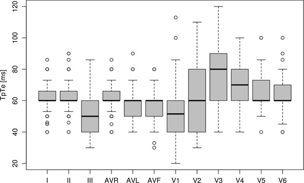
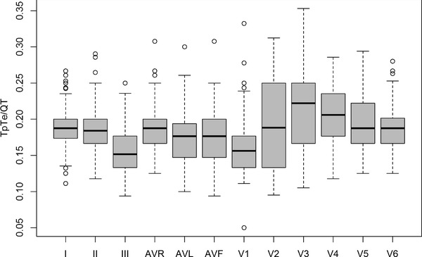
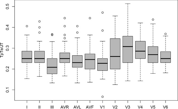
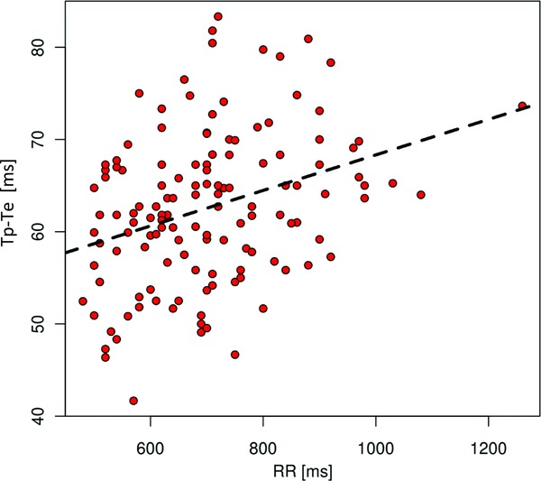
Results: TpTe intervals vary significantly (P < 0.0001) between individual leads-the longest were in lead V3 , the shortest ones in leads III and V1 . Boys had longer TpTe intervals, with statistically significant differences in leads I, aVR and precordial V2 -V6 . Greater values were also observed in older children. TpTe dispersion varied from 6 to 80 ms (mean 38.6 ms ± 14.6 ms, median 40 ms) with no gender differences and greater values in older subjects (P = 0.003). In most leads, higher TpTe/QT and TpTe/JT ratios were seen in boys regardless of age. The TpTe intervals lengthens with lowering heart rate.
Conclusions: In healthy children and adolescents, TpTe intervals vary between individual leads of ECG, with the longest in lead V3 . The TpTe interval is longer in boys and in older children and prolongs as heart rate decelerates. TpTe/QT and TpTe/JT ratios are higher in boys. TpTe interval should be measured in precordial leads.
Keywords: TpTe dispersion; TpTe interval; TpTe/QT and TpTe/JT ratio; children.
©2013, Wiley Periodicals, Inc.
Figures
References
-
- Antzelevitch C. Tpeak‐Tend interval as an index of transmural dispersion of repolarization. Eur J Clin Invest 2001;31:555–557. - PubMed
-
- Watanabe N, Kobayashi Y, Tanno K, et al. Transmural dispersion of repolarization and ventricular tachyarrhythmias. J Electrocardiol 2004;37:191–200. - PubMed
-
- Emori T, Antzelevitch C. Cellular basis for complex T waves and arrhythmic activity following combined I(Kr) and I(Ks) block. J Cardiovasc Electrophysiol 2001;12:1369–1378. - PubMed
MeSH terms
LinkOut - more resources
Full Text Sources
Other Literature Sources
