

An evaluation of meniscal collagenous structure using optical projection tomography
- PMID: 23879345
- PMCID: PMC3726444
- DOI: 10.1186/1471-2342-13-21
An evaluation of meniscal collagenous structure using optical projection tomography
Abstract
Background: The collagenous structure of menisci is a complex network of circumferentially oriented fascicles and interwoven radially oriented tie-fibres. To date, examination of this micro- architecture has been limited to two-dimensional imaging techniques. The purpose of this study was to evaluate the ability of the three-dimensional imaging technique; optical projection tomography (OPT), to visualize the collagenous structure of the meniscus. If successful, this technique would be the first to visualize the macroscopic orientation of collagen fascicles in 3-D in the meniscus and could further refine load bearing mechanisms in the tissue. OPT is an imaging technique capable of imaging samples on the meso-scale (1-10 mm) at a micro-scale resolution. The technique, similar to computed tomography, takes two-dimensional images of objects from incremental angles around the object and reconstructs them using a back projection algorithm to determine three-dimensional structure.
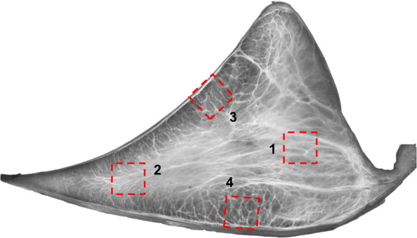
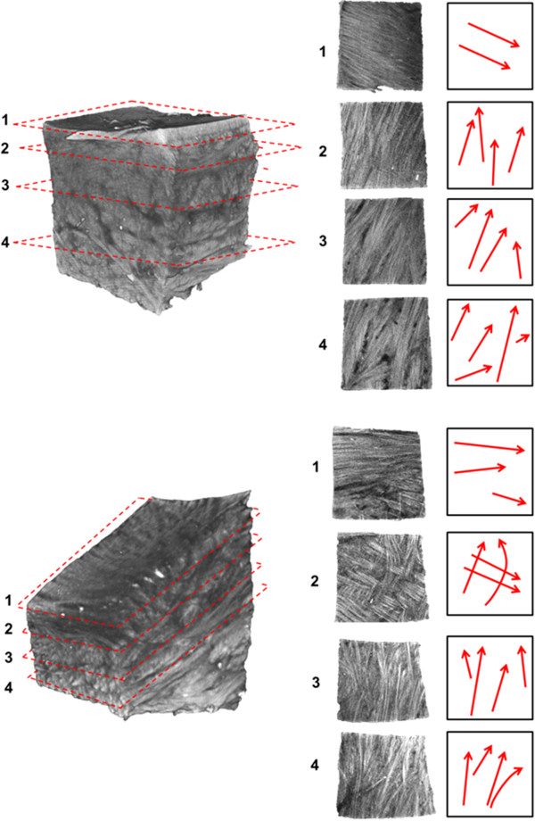
Methods: Bovine meniscal samples were imaged from four locations (outer main body, femoral surface, tibial surface and inner main body) to determine the variation in collagen orientation throughout the tissue. Bovine stifles (n = 2) were obtained from a local abattoir and the menisci carefully dissected. Menisci were fixed in methanol and subsequently cut using a custom cutting jig (n = 4 samples per meniscus). Samples were then mounted in agarose, dehydrated in methanol and subsequently cleared using benzyl alcohol benzyl benzoate (BABB) and imaged using OPT.
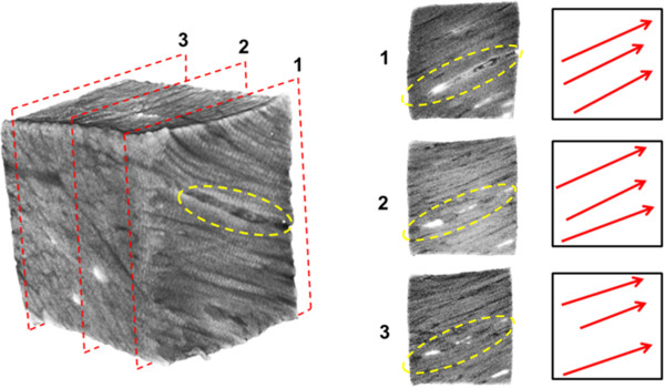
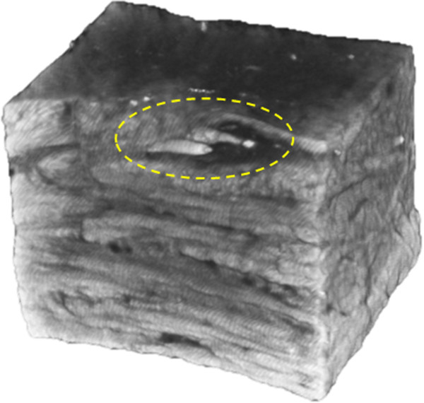
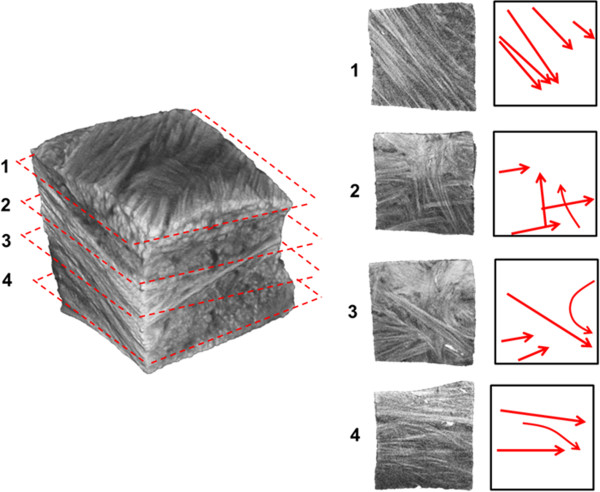
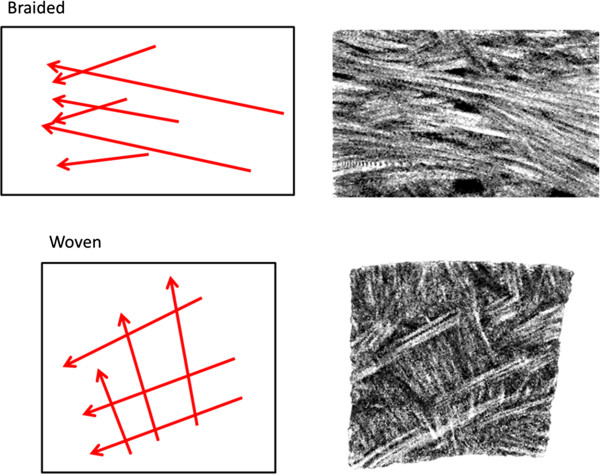
Results: Results indicate circumferential, radial and oblique collagenous orientations at the contact surfaces and in the inner third of the main body of the meniscus. Imaging identified fascicles ranging from 80-420 μm in diameter. Transition zones where fascicles were found to have a woven or braided appearance were also identified. The outer-third of the main body was composed of fascicles oriented predominantly in the circumferential direction. Blood vessels were also visualized using this technique, as their elastin content fluoresces more brightly than collagen at the 425 nm wavelength used by the OPT scanner.
Conclusions: OPT was capable of imaging the collagenous structure, as well as blood vessels in the bovine meniscus. Collagenous structure variability, including transition zones between structural regions not previously described in the meniscus, was identified using this novel technique.
Figures
References
Publication types
MeSH terms
Grants and funding
LinkOut - more resources
Full Text Sources
Other Literature Sources
Medical
Miscellaneous
