

Characterization of a new pathogenic Acanthamoeba Species, A. byersi n. sp., isolated from a human with fatal amoebic encephalitis
- PMID: 23879685
- PMCID: PMC4618466
- DOI: 10.1111/jeu.12069
Characterization of a new pathogenic Acanthamoeba Species, A. byersi n. sp., isolated from a human with fatal amoebic encephalitis
Abstract
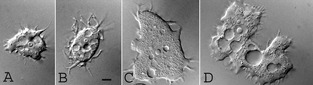
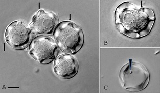
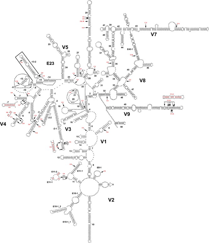
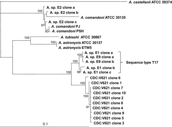
Acanthamoeba spp. are free-living amoebae that are ubiquitous in natural environments. They can cause cutaneous, nasopharyngeal, and disseminated infection, leading to granulomatous amebic encephalitis (GAE) in immunocompromised individuals. In addition, they can cause amoebic keratitis in contact lens wearers. Acanthamoeba GAE is almost always fatal because of difficulty and delay in diagnosis and lack of optimal antimicrobial therapy. Here, we report the description of an unusual strain isolated from skin and brain of a GAE patient. The amoebae displayed large trophozoites and star-shaped cysts, characteristics for acanthamoebas belonging to morphology Group 1. However, its unique morphology and growth characteristics differentiated this new strain from other Group 1 species. DNA sequence analysis, secondary structure prediction, and phylogenetic analysis of the 18S rRNA gene confirmed that this new strain belonged to Group 1, but that it was distinct from the other sequence types within that group. Thus, we hereby propose the establishment of a new species, Acanthamoeba byersi n. sp. as well as a new sequence type, T18, for this new strain. To our knowledge, this is the first report of a Group 1 Acanthamoeba that is indisputably pathogenic in humans.
Keywords: 18S rRNA gene; Group 1 Acanthamoeba; human isolate, nuclear small subunit ribosomal RNA gene; ribosomal secondary structure; sequence type T18.
Published 2013. This article is a U.S. Government work and is in the public domain in the USA.
Figures
References
-
- Amaral‐Zettler, L. A. , Cole, J. , Laatsch, A. D. , Nerad, T. A. , Anderson, O. R. & Reysenbach, A. L. 2006. Vannella epipetala n. sp. isolated from the leaf surface of Spondias mombin (Anacardiaceae) growing in the dry forest of Costa Rica. J. Eukaryot. Microbiol., 53:522–530. - PubMed
-
- Byers, T. J. , Hugo, E. R. & Stewart, V. J. 1990. Genes of Acanthamoeba: DNA, RNA and protein sequences (a review). J. Protozool., 37:17S–25S. - PubMed
-
- Callicott Jr, J. H. 1968. Amebic meningoencephalitis due to free‐living amebas of the Hartmannella (Acanthamoeba)‐Naegleria group. Am. J. Clin. Pathol., 49:84–91. - PubMed
-
- Corsaro, D. & Venditti, D. 2010. Phylogenetic evidence for a new genotype of Acanthamoeba (Amoebozoa, Acanthamoebida). Parasitol. Res., 107:233–238. - PubMed
Publication types
MeSH terms
Substances
Associated data
- Actions
- Actions
- Actions
- Actions
- Actions
- Actions
- Actions
- Actions
- Actions
- Actions
- Actions
- Actions
- Actions
- Actions
Grants and funding
LinkOut - more resources
Full Text Sources
Other Literature Sources
Molecular Biology Databases
Miscellaneous
