. 2013;15(4):R58.
doi: 10.1186/bcr3452.

Development of a screen to identify selective small molecules active against patient-derived metastatic and chemoresistant breast cancer cells

Development of a screen to identify selective small molecules active against patient-derived metastatic and chemoresistant breast cancer cells

Keith M Gligorich et al. Breast Cancer Res. 2013.

Abstract

Introduction: High failure rates of new investigational drugs have impaired the development of breast cancer therapies. One challenge is that excellent activity in preclinical models, such as established cancer cell lines, does not always translate into improved clinical outcomes for patients. New preclinical models, which better replicate clinically-relevant attributes of cancer, such as chemoresistance, metastasis and cellular heterogeneity, may identify novel anti-cancer mechanisms and increase the success of drug development.

Methods: Metastatic breast cancer cells were obtained from pleural effusions of consented patients whose disease had progressed. Normal primary human breast cells were collected from a reduction mammoplasty and immortalized with human telomerase. The patient-derived cells were characterized to determine their cellular heterogeneity and proliferation rate by flow cytometry, while dose response curves were performed for chemotherapies to assess resistance. A screen was developed to measure the differential activity of small molecules on the growth and survival of patient-derived normal breast and metastatic, chemoresistant tumor cells to identify selective anti-cancer compounds. Several hits were identified and validated in dose response assays. One compound, C-6, was further characterized for its effect on cell cycle and cell death in cancer cells.

Results: Patient-derived cells were found to be more heterogeneous, with reduced proliferation rates and enhanced resistance to chemotherapy compared to established cell lines. A screen was subsequently developed that utilized both tumor and normal patient-derived cells. Several compounds were identified, which selectively targeted tumor cells, but not normal cells. Compound C-6 was found to inhibit proliferation and induce cell death in tumor cells via a caspase-independent mechanism.

Conclusions: Short-term culture of patient-derived cells retained more clinically relevant features of breast cancer compared to established cell lines. The low proliferation rate and chemoresistance make patient-derived cells an excellent tool in preclinical drug development.


Figures
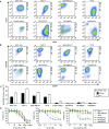
Figure 1
Figure 1
Comparison of established cell lines and patient-derived cells. (A) MCF-10A, MCF-7, MDA-MB-231, T47D, hTERT-HMEC, PE1007070, PE1008032 and PE904557a cells were stained with 7-AAD and a lineage cocktail in combination with CD44/CD24 or (B) CD49f/EPCAM and were analyzed by FACS. The heterogeneity factor (HF) (to the right of each graph) was calculated by multiplying the percent CV of each axis. (C) MCF-10A, MCF-7, MDA-MB-231, T47D, hTERT-HMEC, PE1007070, PE1008032 and PE904557a cells were treated with 10 μM BrdU or EdU for either 30 minutes or six hours and then BrdU/EdU incorporation was analyzed by flow cytometry. The graph shows the percent BrdU/EdU positive cells. (D) Dose response curves of doxorubicin, taxol and gemcitabine against MCF-7, MDA-MB-231, T47D, PE1007070, PE1008032 and PE904557a cells after four days of treatment. Cell viability was measured using a luciferase-based ATP assay and was normalized to the vehicle control. Error bars represent standard deviation. 7-AAD, 7-amino-actinomycin D; BrdU, 5-bromo-2-deoxyuridine; CV, coefficient of variance; EdU, 5-ethynyl-2'-deoxyuridine; FACS, fluorescence activated cell sorting; human mammary epithelial cells; hTERT, human telomerase; PE, pleural effusion.
Figure 2
Figure 2
Identification of the small molecule C-6. (A) PE1007070 and hTERT-HMEC cells were treated with 20 μM of the compound library in duplicate. After four days, a luciferase-based ATP assay was performed and data were normalized to DMSO vehicle control wells to determine viability (%). The difference in viability between the hTERT-HMEC and PE1007070 cells is plotted. The hit limit was 2.7 times the standard deviation. (B) The structure of C-6, which had a 62% difference in viability between the hTERT-HMEC and PE1007070 cells. (C) Dose response curves of C-6 and EC50 values of hTERT-HMEC, PE1007070, PE1008032, and PE904557a cells and (D) MCF-10A, MCF-7, T47D and MDA-MB-231 cells after four days of treatment. N/A indicates data could not be fitted. (E) MCF-10A, MCF-7, MDA-MB-231 and T47D cells were treated with DMSO or 15 μM C-6 for 24 or 48 hours followed by addition of 10 μM BrdU for 30 minutes. The cells were stained for BrdU and propidium iodide and analyzed by flow cytometry. The average ± standard deviation of the percent BrdU positive cells (S phase) of three replicates was calculated. Asterisks (*) denote P-value < 0.05. BrdU, 5-bromo-2-deoxyuridine; DMSO, dimethyl sulfoxide; HMEC, human mammary epithelial cells; hTERT, human telomerase; PE pleural effusion.
Figure 3
Figure 3
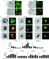
C-6 can induce death in cancer cells. (A) Differential interference contrast (DIC), calcein-AM fluorescence (green), and ethidium homodimer-1 (red) images of hTERT-HMEC and PE1007070 cells cultured in two dimensions and treated with DMSO or 20 μM C-6 for five days (Scale bar is 50 μm). (B) Confocal DIC, calcein-AM fluorescence (green), and ethidium homodimer-1 (red) images of PE904557a, PE900642a, and PE1100025 cells cultured in three-dimensional Matrigel and treated with DMSO or either 30 or 60 μM C-6 for five days (Scale bar is 50 μm). (C) MCF-10A, MCF-7, MDA-MB-231, PE1007070, PE1008032, PE904557a, and PE1100025 cells were treated with DMSO or 30 μM C-6 for 24 to 120 hours. The relative amount of released protease activity was measured using a luciferase-based AAF-Glo assay and these data were normalized to total protein measured with a BCA assay. Asterisks (*) denote P-value < 0.05. DMSO, dimethyl sulfoxide; HMEC, human mammary epithelial cells; hTERT, human telomerase; PE, pleural effusion.
Figure 4
Figure 4
C-6 does not induce caspase or PARP cleavage. (A) MCF-10A, MCF-7, T47D, MDA-MB-231, hTERT-HMEC, PE1007070, PE1008032 and PE904557a cells were treated with DMSO (48 hours), 30 μM C-6 (48 hours), 1 μM staurosporine (24 hours) or 1 μg/mL TRAIL and 5 μM doxorubicin (24 hours) and the resulting whole cell lysates were analyzed by Western blot for cleaved caspase 3, caspase 8, cleaved caspase 9 and PARP. (B) MCF-10A, MCF-7, T47D, MDA-MB-231, PE1007070, PE1008032, PE904557a and PE110025 cells were treated with DMSO (48 hours), 30 μM C-6 (24 and 48 hours) or 1 μM staurosporine (24 hours) and the relative amount of caspase activity was measured using a luciferase-based Caspase-Glo assay and normalized to cell viability using a luciferase-based ATP assay. DMSO, dimethyl sulfoxide; HMEC, human mammary epithelial cells; hTERT, human telomerase; PE, pleural effusion; TRAIL, tumor necrosis factor related apoptosis inducing ligand.

References

    1. Moulder S, Hortobagyi GN. Advances in the treatment of breast cancer. Clin Pharmacol Ther. 2008;15:26–36. doi: 10.1038/sj.clpt.6100449. - DOI - PubMed
    1. Swain SM. Chemotherapy: updates and new perspectives. Oncologist. 2011;15:30–39. doi: 10.1634/theoncologist.2011-S1-30. - DOI - PubMed
    1. DeSantis C, Siegel R, Bandi P, Jemal A. Breast cancer statistics, 2011. CA: Cancer J Clin. 2011;15:408–418. doi: 10.3322/caac.20134. - DOI - PubMed
    1. Gonzalez-Angulo AM, Morales-Vasquez F, Hortobagyi GN. Overview of resistance to systemic therapy in patients with breast cancer. Adv Exp Med Biol. 2007;15:1–22. doi: 10.1007/978-0-387-74039-3_1. - DOI - PubMed
    1. Azim HA, de Azambuja E, Colozza M, Bines J, Piccart MJ. Long-term toxic effects of adjuvant chemotherapy in breast cancer. Ann Oncol. 2011;15:1939–1947. doi: 10.1093/annonc/mdq683. - DOI - PubMed

Publication types

MeSH terms