Review
. 2014 Jun:123:131-40.
doi: 10.1016/j.exer.2013.07.012. Epub 2013 Jul 20.

Regulation of Müller glial dependent neuronal regeneration in the damaged adult zebrafish retina

Affiliations
Review

Regulation of Müller glial dependent neuronal regeneration in the damaged adult zebrafish retina

Ryne A Gorsuch et al. Exp Eye Res. 2014 Jun.

Abstract

This article examines our current knowledge underlying the mechanisms involved in neuronal regeneration in the adult zebrafish retina. Zebrafish, which has the capacity to regenerate a wide variety of tissues and organs (including the fins, kidney, heart, brain, and spinal cord), has become the premier model system to study retinal regeneration due to the robustness and speed of the response and the variety of genetic tools that can be applied to study this question. It is now well documented that retinal damage induces the resident Müller glia to dedifferentiate and reenter the cell cycle to produce neuronal progenitor cells that continue to proliferate, migrate to the damaged retinal layer and differentiate into the missing neuronal cell types. Increasing our understanding of how these cellular events are regulated and occur in response to neuronal damage may provide critical information that can be applied to stimulating a regeneration response in the mammalian retina. In this review, we will focus on the genes/proteins that regulate zebrafish retinal regeneration and will attempt to critically evaluate how these factors may interact to correctly orchestrate the definitive cellular events that occur during regeneration.

Keywords: Ascl1a; Müller glia; Stat3; TNFα; dedifferentiation; neuronal progenitor cells; regeneration; zebrafish.


Figures

Figure 1
Figure 1
Defining the cellular events during regeneration of the light-damaged zebrafish retina. Immunofluorescence of retinal cryosections (A-F) using antibodies to Rhodopsin (Rho; red), PCNA (green), TUNEL (orange) and nuclei (DAPI; blue). Time points correspond to the indicated cellular events, which are summarized in the schematic (G-L). Müller glia are coded as follows: PPMG, red; SPMG, yellow; QMG, green. Neuronal progenitor cells are represented as fusiform cells (cyan). Committed rod and cone progenitors in the ONL are represented as lighter shades of blue and magenta, respectively. Scale bar in panel A is 25 microns and is the same for panels B-F. GCL, Ganglion cell layer; INL, Inner nuclear layer; ONL, Outer nuclear layer; PCNA, Proliferating cell nuclear antigen; PPMG, primary proliferating Müller glia; QMG, quiescent Müller glia; SPMG, secondary proliferating Müller glia.
Figure 2
Figure 2
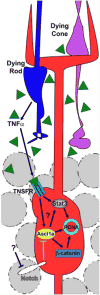
Signaling events necessary for Müller glial dedifferentiation and proliferation. Dying retinal neurons (e.g. rod and cone photoreceptors) secrete TNFα (green triangles), which activates a dedifferentiation program in primary proliferating Müller glia (PPMG). Black lines represent active signaling and grey lines indicate repressed signaling. Dashed objects represent inactive components. Question marks indicate uncertainty between the indicated signaling interactions. The identity of the Notch ligand and its location, either in the photoreceptors or an INL cell type, remain unknown. Ascl1a, Achaete-scute homolog 1a; PCNA, Proliferating cell nuclear antigen: Stat3, Signal transducer and activation of transcription 3; TNFα, Tumor necrosis factor-alpha; TNFSR, TNF superfamily receptor.
Figure 3
Figure 3
Recruitment of SPMG and maintenance of QMG. PPMG secrete TNFα (green triangles) in a Stat3- and Ascl1a-dependent manner, and HB-EGF (purple circles), It is not entirely clear what stimulates HB-EGF expression, although Ascl1a may be involved. These secreted factors bind their respective receptors in neighboring Müller glia, which activates a secondary dedifferentiation program (SPMG). A concentration gradient of TNFα and HB-EGF likely activates neighboring Müller glia at a certain threshold, below which Müller glia do not dedifferentiate and remain quiescent (QMG). Inhibition of Notch signaling results in dedifferentiation of QMG (Wan et al., 2012), suggesting Notch expression may be negatively correlated to the TNFα/HB-EGF gradient. Because Ascl1a can canonically activate Delta ligands, Delta expression in PPMG/SPMG may reinforce Notch expression in neighboring Müller glia outside of the TNFα/HB-EGF gradient. Black lines indicate active signaling, while grey lines represent repressed signaling. Dashed objects represent inactive components. While Notch expression/activity is likely repressed in the SPMG, the identity and location, either the photoreceptors and/or INL cell type, of the corresponding ligand remains unknown. Question marks indicate uncertainty between the specified signaling interactions. Ascl1a, Achaete-scute homolog 1a; EGFR, epidermal growth factor receptor; ERK1/2, Extracellular signal-regulated kinase 1 and 2; HB-EGF, Heparin-binding EGF-like growth factor; Insm1a, Insulinoma-associated 1a; PCNA, Proliferating cell nuclear antigen; PPMG, primary proliferating Müller glia; QMG, Quiescent Müller glia; SPMG, Secondary proliferating Müller glia; Stat3, Signal activator and transducer of transcription 3; TNFα, Tumor necrosis factor-alpha; TNFSR, TNF superfamily receptor.

References

    1. Agathocleous M, Iordanova I, Willardsen MI, Xue XY, Vetter ML, Harris WA, Moore KB. A directional Wnt/beta-catenin-Sox2-proneural pathway regulates the transition from proliferation to differentiation in the Xenopus retina. Development. 2009;136:3289–3299. - PMC - PubMed
    1. Bernardos RL, Barthel LK, Meyers JR, Raymond PA. Late-stage neuronal progenitors in the retina are radial Muller glia that function as retinal stem cells. J Neurosci. 2007;27:7028–7040. - PMC - PubMed
    1. Bernardos RL, Lentz SI, Wolfe MS, Raymond PA. Notch-Delta signaling is required for spatial patterning and Muller glia differentiation in the zebrafish retina. Dev Biol. 2005;278:381–395. - PubMed
    1. Braunger BM, Ohlmann A, Koch M, Tanimoto N, Volz C, Yang Y, Bosl MR, Cvekl A, Jagle H, Seeliger MW, Tamm ER. Constitutive overexpression of Norrin activates Wnt/beta-catenin and endothelin-2 signaling to protect photoreceptors from light damage. Neurobiol Dis. 2013;50:1–12. - PubMed
    1. Calinescu AA, Vihtelic TS, Hyde DR, Hitchcock PF. Cellular expression of midkine-a and midkine-b during retinal development and photoreceptor regeneration in zebrafish. J Comp Neurol. 2009;514:1–10. - PMC - PubMed

Publication types