

A novel hybrid CFHR1/CFH gene causes atypical hemolytic uremic syndrome
- PMID: 23880784
- PMCID: PMC4433496
- DOI: 10.1007/s00467-013-2560-2
A novel hybrid CFHR1/CFH gene causes atypical hemolytic uremic syndrome
Abstract
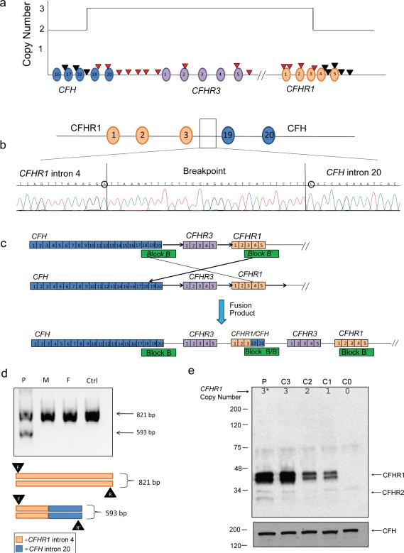
Background: Mutations in complement factor H (CFH) are associated with complement dysregulation and the development of an aggressive form of atypical hemolytic uremic syndrome (aHUS) that progresses to end-stage renal disease (ESRD) and in most patients has a high rate of recurrence following transplantation. Sequence analysis of CFH and its downstream complement factor H-related genes (CFHR1-5) reveals several macrohomologous blocks caused by large genomic duplications. This high degree of sequence identity renders this area susceptible to nonallelic homologous recombination (NAHR) events, resulting in large-scale deletions, duplications, and the generation of hybrid CFH genes.
Case-diagnosis: Here, we report the finding of a novel CFHR1/CFH hybrid gene created by a de novo NAHR event in a 14-year-old girl with aHUS. The resulting fusion protein contains the first three short consensus repeats (SCRs) of CFHR1 and the terminal two SCRs of CFH.
Conclusions: This finding demonstrates a novel pathogenic mechanism for the development of aHUS. Additionally, since standard Sanger sequencing is unable to detect such rearrangements, all aHUS patients should receive comprehensive genetic screening that includes analysis of copy number variation in order to identify patients with poor clinical prognoses.
Figures
References
-
- Maga TK, Nishimura CJ, Weaver AE, Frees KL, Smith RJH. Mutations in alternative pathway complement proteins in American patients with atypical hemolytic uremic syndrome. Hum Mutat. 2010;31:E1445–E1460. - PubMed
-
- Sellier-Leclerc A, Fremeaux-Bacchi V, Dragon-Durey M, Macher M-A, Niaudet P, Guest G, Boudailliez B, Bouissou F, Deschenes G, Gie S, Tsimaratos M, Fischbach M, Morin D, Nivet H, Alberti C, Loirat C. Differential impact of complement mutations on clinical characteristics in atypical hemolytic uremic syndrome. J Am Soc Nephrol. 2007;18:2392–2400. - PubMed
-
- Caprioli J, Noris M, Brioschi S, Pianetti G, Castelletti F, Bettinaglio P, Mele C, Bresin E, Cassis L, Gamba S, Porrati F, Bucchioni S, Monteferrante G, Fang CJ, Liszewski MK, Kavanagh D, Atkinson JP, Remuzzi G. Genetics of HUS: the impact of MCP, CFH, and IF mutations on clinical presentation, response to treatment, and outcome. Blood. 2006;108:1267–1279. - PMC - PubMed
-
- Józsi M, Zipfel PF. Factor H family proteins and human diseases. Trends Immunol. 2008;29:380–387. - PubMed
Publication types
MeSH terms
Substances
Grants and funding
LinkOut - more resources
Full Text Sources
Other Literature Sources
Miscellaneous
