. 2013 Oct;4(5):527-38.
doi: 10.1007/s13244-012-0211-y. Epub 2013 Jul 25.

Breast disease in the pregnant and lactating patient: radiological-pathological correlation

Affiliations

Breast disease in the pregnant and lactating patient: radiological-pathological correlation

Surekha Joshi et al. Insights Imaging. 2013 Oct.

Abstract

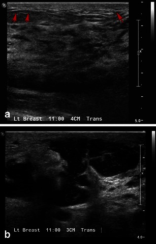
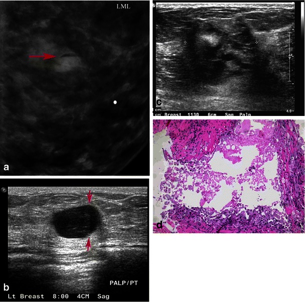
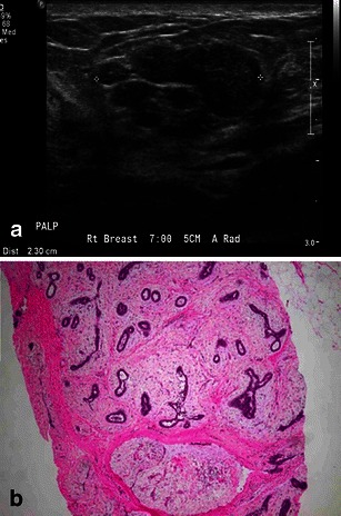
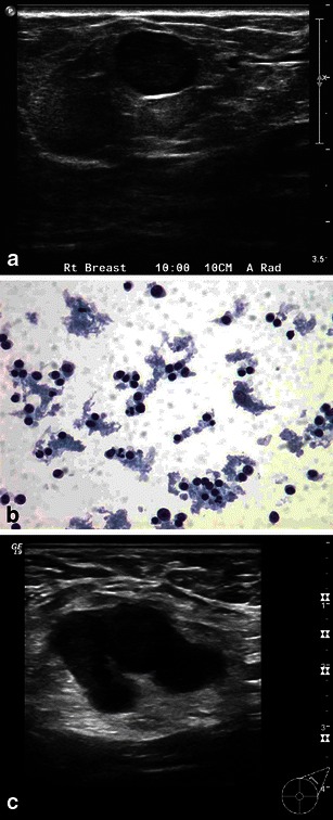
Objective: Substantial physiological changes occur during pregnancy and lactation, making breast evaluation challenging in these patients. This article reviews the imaging challenges of the breast during pregnancy and lactation. The normal imaging appearance, imaging protocols and the imaging features of each commonly encountered benign and malignant entity with pathological correlation and supporting examples is described. An awareness of the imaging features of the breast during these physiological states and of various benign and malignant diseases that occur permits optimal management.

Conclusions: Evaluation of the pregnant and lactating patients who present with a breast problem is challenging. Although ultrasound may characterise the finding in many cases, mammography and even MRI may have a role in the management of these patients.

Teaching points: • To review physiological changes of the breast during pregnancy and lactation • To review imaging protocols of the breast during pregnancy and lactation • Discuss imaging findings with pathological correlation of benign and malignant diseases in pregnancy and lactation • Discuss pathological correlation of imaging findings in pregnancy and lactation.


Figures

Fig. 1
Fig. 1
Normal pregnancy and lactational change: a 39-year-old woman, lactating for 3 months. a Note the larger breast size and increased density of parenchyma in spite of pumping prior to imaging on right MLO view compared with b, reduced density of parenchyma on mammogram performed prior to pregnancy. c Pregnancy change: a 38-year-old woman, 24 weeks pregnant presents with breast pain. Note diffuse hypoechogenicity due to enlargement of non-fatty fibroglandular components during pregnancy. d Increased echogenicity with ductal prominence, which is typical appearance of a lactating breast on grey-scale ultrasound
Fig. 2
Fig. 2
a Normal lactating tissue showing diffuse rapid enhancement on dynamic contrast enhanced MRI. b Note the bright ducts (black arrows) on T2- weighted images
Fig. 3
Fig. 3
a A 44-year-old woman, 10 months postpartum and lactating. Note the irregular spiculated mass (white arrows) showing low signal intensity on T2–weighted images and (b) intense enhancement (white arrows) on dynamic contrast enhanced mages. This was a biopsy proven invasive lobular carcinoma with lymphovascular invasion
Fig. 4
Fig. 4
a A 36-year-old woman, 12 months postpartum and lactating. Note the mass (white arrows) showing high signal intensity on T2–weighted images and (b) increased enhancement (white arrows) on dynamic contrast-enhanced images, compared with the rest of the breast tissue. This was a biopsy-proven grade 3, invasive ductal carcinoma
Fig. 5
Fig. 5
Lactational change: a 31-year-old lactating woman, presenting with lump in the right breast. a Grey-scale ultrasound shows a partially circumscribed hypoechoic nodule with cystic areas, (b) with internal vascularity on power Doppler. c Pathology slide at high power (20×) shows lobular expansion containing increased numbers of acini, many of which are enlarged and dilated, consistent with lactational change
Fig. 6
Fig. 6
Lactating adenoma: a 38-year-old pregnant woman, presenting with a breast lump. a Grey-scale ultrasound image shows an isoechoic, circumscribed nodule. b Power Doppler image shows minimal peripheral vascularity. Biopsy was performed revealing lactating adenoma. c Higher-power image (20×) shows epithelial cell enlargement, cytoplasmic vacuolisation and a hobnail appearance with protrusion of cells into the acinar lumen
Fig. 7
Fig. 7
Lactational calcifications. a A 39-year-old lactating woman with an area of palpable concern in the right breast. A mammogram of the left breast, MLO view, shows incidental new diffuse microcalcifications (black arrows). Similar findings were also appreciated in the opposite breast. All of these calcifications represent lactational deposits. b A 38-year-old lactating woman presented with a right breast lump; incidental focal microcalcifications (black arrows) were seen in the left breast at 12:00, magnification view in CC and ML view (not shown here) showed a focal cluster of slightly pleomorphic calcifications. Stereotactic biopsy was performed, which revealed lactational calcifications
Fig. 8
Fig. 8
Blocked milk duct: a 35-year-old lactating woman with fluctuant non-tender breast mass. a Grey-scale ultrasound image reveals linear hypoechoic structure with no vascularity on power Doppler (not shown here). Findings were consistent with a blocked milk duct. b A 27-year-old lactating woman, presenting with tender mass in left axilla. Grey-scale ultrasound reveals a hypoechoic tubular lesion with no vascularity on power Doppler (not shown here). This was aspirated, for symptomatic relief to the patient, yielding milky fluid
Fig. 9
Fig. 9
a Mastitis: a 29-year-old lactating woman, presenting with breast pain and erythema. Ultrasound reveals skin thickening and no discrete abscess consistent with mastitis. Patient’s redness and pain resolved after treatment with antibiotics. b Breast abscess: a 39-year-old lactating woman with left breast erythema swelling and fever. Grey-scale ultrasound shows a multiloculated fluid collection with debris representing abscess. Aspiration yielded pus, and microbiology revealed MRSA
Fig. 10
Fig. 10
Galactocele: a 35-year-old lactating woman, presenting with a palpable lump. a Photographic magnification of left lateral mammogram shows a well-circumscribed lesion with a fat fluid level on the lateral projection. This is the classic appearance of galactocele. b Grey-scale ultrasound shows a cystic lesion which, on adjusting the gain, shows a fat/fluid level diagnostic of galactocele. c Another patient, a 38-year-old lactating woman, presenting with breast pain and palpable mass. Mammogram showed dense breast tissue with a partially obscured mass and skin thickening (not shown here). Grey-scale ultrasound image shows heterogeneous ill-defined mass, which was vascular on power Doppler. Biopsy was performed. d Microscopic high-power image (40×), revealing a ruptured galactocele with cyst contents leaking into the surrounding tissue, causing a lipogranulomatous inflammation and foamy histiocyte aggregation
Fig. 11
Fig. 11
Fibroadenoma: a 22-year-old pregnant woman, presenting with a lump. a Grey-scale ultrasound shows a partially circumscribed macrolobulated nodule without internal vascularity. Core-needle biopsy was performed. b Microscopic low power (10×) reveals a fibroadenoma, illustrating benign proliferation of glands and stroma
Fig. 12
Fig. 12
a Benign reactive lymph node: a 30-year-old pregnant woman, presenting with a palpable lump. Grey-scale ultrasound image shows well circumscribed hypoechoic nodule, with significant vascularity on power Doppler. b Fine-needle aspiration cytology reveals polymorphous population of lymphocytes and macrophages, all features of benign reactive lymph node. c Metastatic lymph node: a 39-year-old woman, 12 weeks pregnant, presenting with palpable axillary mass. The patient had a remote history of breast cancer treated with mastectomy. Gray-scale ultrasound showed an enlarged axillary node with abnormally thickened cortex. Core biopsy revealed metastatic breast cancer
Fig. 13
Fig. 13
Granulomatous mastitis: a 30-year-old woman, postpartum, presenting with erythema, skin thickening and draining sinus. Grey-scale ultrasound image (a, b) showed irregular hypoechoic mass with peripheral vascularity. Biopsy was performed; high power image (40×) (c) showed non-necrotising granulomas, lymphocytes and plasma cells in the background. Special stains for fungus and mycobacterium were negative. Follow-up after conservative treatment and steroids were done. The follow-up ultrasound image (d) revealed significant improvement. Findings were consistent with granulomatous mastitis
Fig. 14
Fig. 14
Pregnancy-associated breast cancer: a 36-year-old woman, 19 weeks pregnant, presenting with a palpable lump. Mammogram (a, b) reveals a dense partially circumscribed mass in the upper outer quadrant with associated microcalcifications. c Grey-scale ultrasound shows a hypoechoic ill-defined mass with hyperechoic foci of microcalcifications. d Power Doppler shows internal vascularity. e Microscopic high-power image (40×) shows grade 3 invasive ductal carcinoma with lack of tubule formation, pleomorphic nuclei and abundant mitotic figures

References

    1. Sabate JM, Clotet M, Torrubia S, et al. Radiologic evaluation of breast disorders related to pregnancy and lactation. Radiographics. 2007;27(Suppl 1):S101–S124. doi: 10.1148/rg.27si075505. - DOI - PubMed
    1. Robbins J, Jeffries D, Roubidoux M, Helvie M (2011) Accuracy of diagnostic mammography and breast ultrasound during pregnancy and lactation. AJR Am J Roentgenol 196:716–722 - PubMed
    1. Magno S, Terribile D, Franceschini G, et al. Early onset lactating adenoma and the role of breast MRI: a case report. J Med Case Rep. 2009;3:43. doi: 10.1186/1752-1947-3-43. - DOI - PMC - PubMed
    1. National Comprehensive Cancer Network (2008) Clinical practice guidelines in oncology: breast cancer. In: National comprehensive cancer network, Vol. 2. NCCN, Fort Washington
    1. National Comprehensive Cancer Network(2008) Clinical practice guidelines in oncology: breast cancer screening and diagnosis guidelines. In: National comprehensive cancer network, Vol. 1. NCCN, Fort Washington - PubMed

LinkOut - more resources