. 2014 Mar;7(2):335-47.
doi: 10.1038/mi.2013.52. Epub 2013 Jul 24.

Central cholinergic activation of a vagus nerve-to-spleen circuit alleviates experimental colitis

Affiliations

Central cholinergic activation of a vagus nerve-to-spleen circuit alleviates experimental colitis

H Ji et al. Mucosal Immunol. 2014 Mar.

Abstract

The cholinergic anti-inflammatory pathway is an efferent vagus nerve-based mechanism that regulates immune responses and cytokine production through α7 nicotinic acetylcholine receptor (α7nAChR) signaling. Decreased efferent vagus nerve activity is observed in inflammatory bowel disease. We determined whether central activation of this pathway alters inflammation in mice with colitis and the mediating role of a vagus nerve-to-spleen circuit and α7nAChR signaling. Two experimental models of colitis were used in C57BL/6 mice. Central cholinergic activation induced by the acetylcholinesterase inhibitor galantamine or a muscarinic acetylcholine receptor agonist treatments resulted in reduced mucosal inflammation associated with decreased major histocompatibility complex II level and pro-inflammatory cytokine secretion by splenic CD11c⁺ cells mediated by α7nAChR signaling. The cholinergic anti-inflammatory efficacy was abolished in mice with vagotomy, splenic neurectomy, or splenectomy. In conclusion, central cholinergic activation of a vagus nerve-to-spleen circuit controls intestinal inflammation and this regulation can be explored to develop novel therapeutic strategies.


Figures

Figure 1
Figure 1. Galantamine (GAL) alleviates the severity of dextran sulphate sodium (DSS)-induced colitis
GAL (6 days, i.p.) treatment was started one day prior to colitis induction. A: Disease activity index; B: Macroscopic scores; C: Colonic myeloperoxidase (MPO) activity; D: Serum C-reactive protein (CRP). Values are shown as means±SEM. Samples were collected on day 5 post-DSS; mice per group ≥8. aP<0.05 as compared to saline DSS-treated group, bP<0.05 as compared to control (H2O)-treated group.
Figure 2
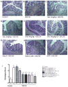
Figure 2. Centrally-acting acetylcholinesterase inhibitors and mAChR ligands alleviate the severity of dextran sulphate sodium (DSS)-induced colitis
Appearance of a colon in mice A: in absence of colitis (control group); B: in mice with DSS-induced colitis; C: in galantamine (GAL) (4mg/kg, i.p. for 6 days)-treated mice with DSS-induced colitis D: in Huperzine A (Hup A) (0.4mg/kg, i.p. for 6 days)-treated mice with DSS-induced colitis E: in McN-A-343 (M1mAChR agonist) (5 ng/kg/day, i.c.v., for 6 days)-treated mice with DSS-induced colitis F: in methoctramine (MTT, M2mAChR antagonist) (5 ng/kg/day, i.c.v., for 6 days)-treated mice with DSS-induced colitis G: Histological score; values are shown as means±SEM. Samples were collected on day 5 post-DSS; mice per group ≥8. aP<0.05 as compared to saline DSS-treated group, bP<0.05 as compared to control (H2O)-group. Hematoxylin and eosin staining, 100X magnifications.
Figure 3
Figure 3. Galantamine (GAL) effects in dextran sulphate sodium (DSS)-induced colitis are mediated through central mAChRs
Atropine methyl nitrate (AMN, a mAChR antagonist that does not cross the blood-brain barrier) (4mg/kg, i.p. for 6 days) or atropine sulfate (AS, a mAChR antagonist that crosses the blood-brain barrier) were administered as a single daily injection 20 min prior to every GAL administration (4mg/kg, i.p., for 6 days). A: Macroscopic score; B: Colonic myeloperoxidase (MPO) activity; C: Serum C-reactive protein (CRP); D: Colonic Interleukin (IL)-1β amount; E: Colonic IL-6 amount; F: Colonic tumor necrosis factor (TNF)-α amount. Values are shown as means±SEM. Samples were collected on day 5 post-DSS; mice per group ≥8. aP<0.05, compared to non DSS-treated group (control, H20) respectively, bP<0.05 as compared to vehicle DSS-treated group respectively, cP<0.05 as compared to GAL-DSS-treated group.
Figure 4
Figure 4. Central administration of a M1mAChR agonist or a M2mAChR antagonist alleviates the severity of dextran sulphate sodium (DSS)-induced colitis
McN-A-343 (M1mAChR agonist) or methoctramine (MTT, M2mAChR antagonist) (5 ng/kg/day, i.c.v., for 6 days) treatments were started one day before induction of colitis. A: Macroscopic score; B: Colonic myeloperoxidase (MPO) activity; C: Serum C-reactive protein (CRP); D: Colonic Interleukin (IL)-1β amount; E: Colonic IL-6 amount; F: Colonic tumor necrosis factor (TNF)-α amount. Values are shown as means±SEM. Samples were collected on day 5 post-DSS; mice per group ≥8. aP<0.05, compared to non DSS-treated group (control, H20) respectively, bP<0.05 as compared to vehicle DSS-treated group.
Figure 5
Figure 5. Galantamine’s effects in mice with dextran sulphate sodium (DSS)-induced colitis are mediated through vagus nerve and splenic nerve signaling to the spleen
Vagotomy (VXP) and/or splenectomy (SPX), splenic neurectomy (NRX) and/or splenectomy (SPX) were performed 10 days prior to initiating galantamine (4 mg/kg/day, i.p.) treatment and/or colitis induction as described in Material and Methods. *Sham represents data obtained in sham SPX mice, because no significant differences were determined between this group and any other sham group of animals; A: Macroscopic score; B: Colonic myeloperoxidase (MPO) activity; C: Serum C-reactive protein (CRP); D: Colonic Interleukin (IL)-1β amount; E: Colonic IL-6 amount; F: Colonic tumor necrosis factor (TNF)-α amount. Values are shown as means±SEM. Samples were collected on day 5 post-DSS; mice per group ≥8. aP<0.05 as compared to sham-saline-DSS-treated group, bP<0.05 as compared to VXP-DSS-treated group or NRX-DSS-treated group, cP<0.05 as compared to sham-GAL-DSS-treated group.
Figure 6
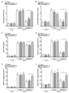
Figure 6. Cholinergic treatments with galantamine (GAL) or McN-A-343 result in increased splenic acetylcholine (ACh) levels, mediated through vagus nerve and splenic nerve signaling
Vagotomy (VXP) and/or splenic neurectomy (NRX) were performed 10 days prior to GAL (4mg/kg/day, i.p. for 6 days) or McN-A-343 (5ng/kg/day, i.c.v., for 6 days) treatments and splenic ACh levels were analyzed as described in Material and Methods. Splenic ACh levels were determined on day 5 post-colitis induction with dextran sulphate sodium (DSS). In colitic control condition, the level of ACh was 1845±85μM/spleen. Values are shown as means±SEM, 3 independent experiments with 4 mice per group. aP<0.05 as compared to control sham-DSS-treated group, bP<0.05 as compared to sham GAL-DSS-treated group, cP<0.05 as compared to sham MCN-A-343-DSS-treated group.
Figure 7
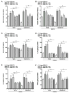
Figure 7. Effects of cholinergic treatment on splenic CD11c+ cells cytokine production in the context of dextran sulphate sodium (DSS)-induced colitis
A: Interleukin (IL)-12p40, IL-1β and IL-6 production from dendritic cells. Splenic CD11c+ cells were isolated from galantamine (GAL, 4 mg/kg/day, i.p. for 6 days)-treated groups of colitic mice subjected to sham-operation, vagotomy (VXP) or splenic neurectomy (NRX) on day 5 post-colitis induction. Splenic CD11c+ cells were also isolated from groups of colitic mice subjected to sham-operation, vagotomy (VXP) or splenic neurectomy (NRX) and incubated ex vivo with GTS-21 (a specific α7nAChR agonist, 100 μM). IL-12p40, IL-1β and IL-6 was measured in media at 24h following treatments. Values are shown as means±SEM, 3 independent experiments with 4 mice per group. aP<0.05 as compared to DSS control group, bP<0.05, n=8.
Figure 8
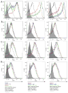
Figure 8. Effect of cholinergic treatments with galantamine (GAL), vagotomy and neurectomy on splenic CD11c+ cells phenotype in the context of dextran sulphate sodium (DSS)-induced colitis
Using CD11c+ MACS positive selection, splenic CD11c+ cells were isolated from galantamine (GAL, 4 mg/kg/day, i.p. for 6 days)-treated or vehicle groups of colitic mice subjected to sham-operation, vagotomy (VXP) or splenic neurectomy (NRX) on day 5 post-colitis induction. Splenic CD11c+ cell phenotype as characterized by median fluorescence intensity (% of Max) A: MHC II (Alexa-647), B: CD40 (FITC), C: CD80 (PE) and D: CD86 (V450) surface expression. Representative results from n=4 per group are shown.

References

    1. Molodecky NA, Soon IS, Rabi DM, Ghali WA, Ferris M, Chernoff G, et al. Increasing incidence and prevalence of the inflammatory bowel diseases with time, based on systematic review. Gastroenterology. 2012;142:46–54. e42. quiz e30. - PubMed
    1. Strober W, Fuss IJ. Proinflammatory cytokines in the pathogenesis of inflammatory bowel diseases. Gastroenterology. 2011;140:1756–1767. - PMC - PubMed
    1. Perrier C, Rutgeerts P. Cytokine blockade in inflammatory bowel diseases. Immunotherapy. 2011;3:1341–1352. - PubMed
    1. Ghia JE, Blennerhassett P, Kumar-Ondiveeran H, Verdu EF, Collins SM. The vagus nerve: a tonic inhibitory influence associated with inflammatory bowel disease in a murine model. Gastroenterology. 2006;131:1122–1130. - PubMed
    1. Goldstein RS, Bruchfeld A, Yang L, Qureshi AR, Gallowitsch-Puerta M, Patel NB, et al. Cholinergic anti-inflammatory pathway activity and High Mobility Group Box-1 (HMGB1) serum levels in patients with rheumatoid arthritis. Mol Med. 2007;13:210–215. - PMC - PubMed

Publication types

MeSH terms