. 2013:3:2271.
doi: 10.1038/srep02271.

Polyreactive antibodies plus complement enhance the phagocytosis of cells made apoptotic by UV-light or HIV

Affiliations

Polyreactive antibodies plus complement enhance the phagocytosis of cells made apoptotic by UV-light or HIV

Zhao-hua Zhou et al. Sci Rep. 2013.

Abstract

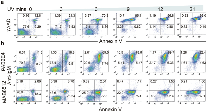
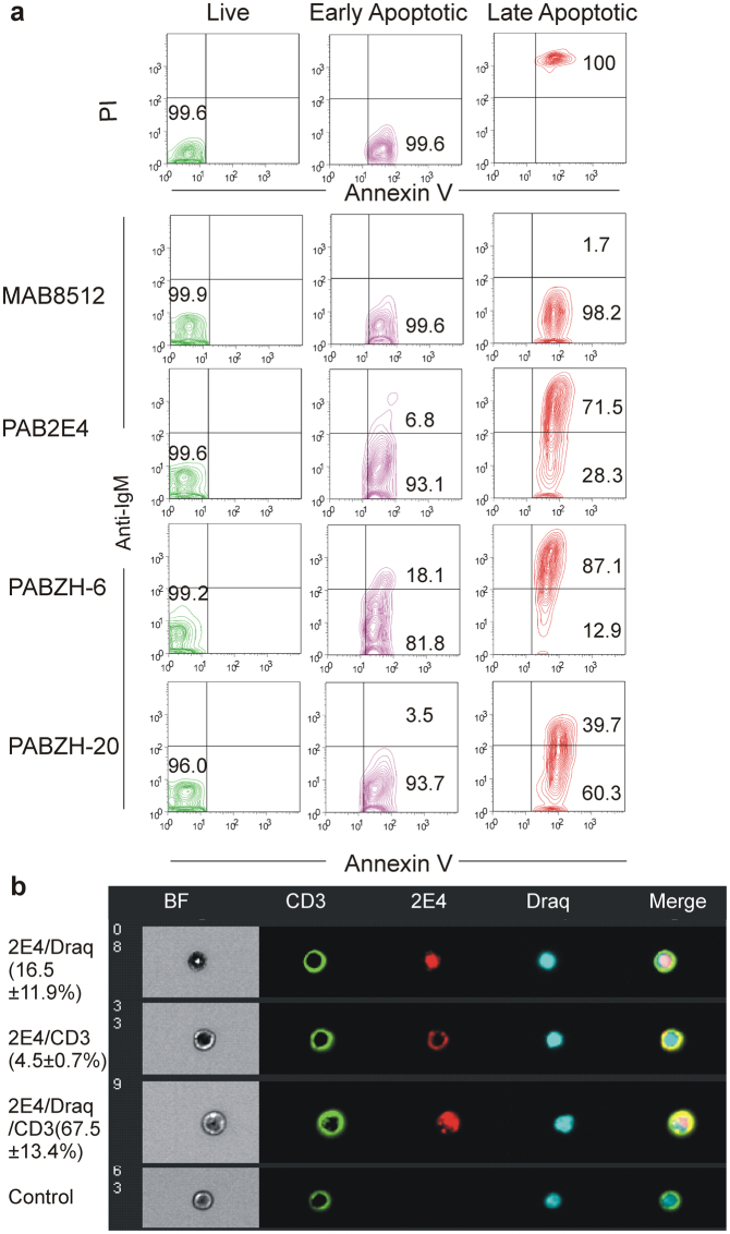
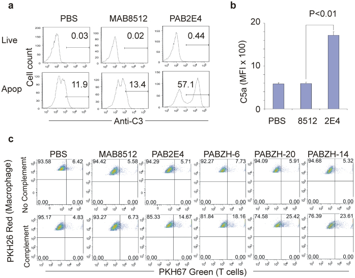
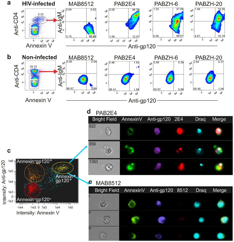
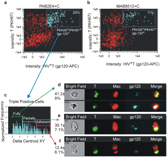
Polyreactive antibodies are a major component of the natural antibody repertoire and are capable of binding a variety of structurally unrelated antigens. Many of the properties attributed to natural antibodies, in fact, are turning out to be due to polyreactive antibodies. In humans, each day, billions of cells undergo apoptosis. In the present experiments, we show by ImageStream technology that although polyreactive antibodies do not bind to live T cells they bind to both the plasma membrane and cytoplasm of late apoptotic cells, fix complement, generate the anaphylatoxin C5a and increase by as much as 5 fold complement-mediated phagocytosis by macrophages. Of particular importance, T cells undergoing apoptosis following infection with HIV also bind polyreactive antibodies and are phagocytosed. We conclude that the polyreactive antibodies in the natural antibody repertoire contribute in a major way to the clearance of cells made apoptotic by a variety of natural and infectious processes.


Conflict of interest statement

The authors declare no competing financial interests.

Figures

Figure 1
Figure 1
Polyreactive antibody 2E4 binds to UV-induced apoptotic T cells. Human T cells were incubated with polyreactive 2E4 or monoreactive 8512 after being exposed to UV light for different lengths of time. (a) Apoptosis as evaluated by the percentage of cells binding Annexin V and 7AAD. (b) Polyreactive 2E4, but not monoreactive 8512, binds to apoptotic cells. Data are representative of three independent experiments with T cells from three different donors.
Figure 2
Figure 2
Polyreactive antibodies bind primarily to late apoptotic cells and to multiple sites on and within apoptotic T cells. (a) Human T cells exposed to UV light were sorted into live (Annexin V/PI), early (Annexin V+/PI) and late (Annexin V+/PI+) apoptotic populations. The binding profile of monoreactive 8512 and polyreactive 2E4, ZH-6 and ZH-20 antibodies showed that the polyreactive antibodies bound primarily to late apoptotic cells. (b) UV-induced late apoptotic human T cells were incubated with polyreactive 2E4 and then stained with FITC-labeled antibody to CD3 (green) to identify the plasma membrane and Draq 5 (blue) to identify the nucleus. PE-labeled anti-immunoglobulin (red) was used to co-localize polyreactive 2E4 with these structures based upon bright field similarity analysis by ImageStream. 2E4/Draq 5, 2E4/CD3 and the 2E4/Draq 5/CD3 co-localization was quantified based on the fraction of each population present in a total of 10,000 cell images obtained from each of the samples collected. Representative images show co-localization (merge) of 2E4 with nucleus (Draq 5: row 1); 2E4 with plasma membrane (CD3: row 2); and 2E4 with cytoplasm, nucleus and plasma membrane (Draq 5 and CD3: row 3). Controls show staining in the absence of 2E4 (row 4). Experiments (panels a and b) were repeated at least three times and each time with T cells from different donors.
Figure 3
Figure 3
Binding of polyreactive antibodies to human apoptotic T cells fixes complement, generates C5a and enhances phagocytosis by macrophages. (a) Apoptotic, but not live, T cells fix complement in the presence of polyreactive 2E4, but not monoreactive 8512. Complement binding was determined by antibody to C3. (b) Apoptotic T cells treated with polyreactive 2E4, but not monoreactive 8512, fix complement and generate the anaphylatoxin C5a as determined by mean fluorescence intensity using BD Cytometric Bead Array. (c) In the presence of complement, there is a 3.0 to 5.0 fold increase in the phagocytosis of apoptotic T cells treated with polyreactive antibodies 2E4, ZH-6, ZH-20 and ZH-14 as compared to cells treated with monoreactive antibody 8512 or PBS. In the absence of complement, there is no increase in phagocytosis over background activity. FACS analysis shows gated PKH26 macrophages. PKH67 single positive T cells were excluded in the analysis.
Figure 4
Figure 4
In vivo phagocytosis of apoptotic thymic T cells treated with polyreactive 2E4 plus complement or monoreactive 8512 plus complement. IgM-deficient mice were injected with PKH26 solution to stain peritoneal macrophages and 24 hours later injected with apoptotic thymic T cells that had been stained with PKH67. Analysis by ImageStream of gated peritoneal macrophages showed (a) that nearly twice as many of the macrophages phagocytosed thymic T cells (PKH26+/PKH67+) that had been exposed to polyreactive 2E4, as compared to (b) thymic T cells that had been exposed to monoreactive 8512. Gating was based upon the negative control, i.e., PKH26-stained macrophages without exposure to PKH67-stained thymic cells. (c) PKH26+/PKH67+ cells from Figure 4a then were divided into fully phagocytosed, partially phagocytosed and adherent cells based upon Delta Centroid XY analysis. (d–f) Representative images taken by ImageStream showing phagocytosis (merge) of apoptotic thymic T cells (green) by macrophages (red): (d) fully phagocytosed; (e) partially phagocytosed; and (f) macrophage/thymic T cell adherence. Data are representative of at least three different experiments.
Figure 5
Figure 5
Polyreactive antibodies bind to HIV-induced apoptotic cells. HIV-infected CD4+ T cells were gated for apoptosis as determined by Annexin V binding. (a) Polyreactive antibodies 2E4, ZH-6 and ZH-20, but not monoreactive antibody 8512, bound to the HIV-infected apoptotic T cells. (b) Neither monoreactive nor polyreactive antibodies bound to the non-infected cells. (c) Human T cells gated by ImageStream into Annexin V/gp120+, Annexin V+/gp120+ and double negative cells. (d) Representative images showing that Annexin V+/gp120+ apoptotic cells bind polyreactive 2E4 antibody, but not (e) monoreactive 8512 antibody.
Figure 6
Figure 6
Phagocytosis of HIV-induced apoptotic T cells by macrophages as determined by ImageStream. Analysis by ImageStream of macrophages showing (a) that nearly twice as many of the HIV-infected (gp120+)T cells that had been exposed to polyreactive 2E4 and complement, as compared to (b) monoreactive 8512 and complement, were phagocytosed by macrophages. Macrophages were gated based upon PKH26 positivity and the PKH26+/PKH67+/gp120+ population was analyzed. (c) Polyreactive 2E4-treated HIV infected T cells (PKH67+/gp120+) phagocytosed by macrophages (PKH26+) were analyzed based upon Delta Centroid XY analysis. (d–f) Representative images showing phagocytosis (merge) of HIV infected T cells (green and APC+) by macrophages (red): (d) fully phagocytosed; (e) partially phagocytosed; and (f) macrophage/T cell adherence. Data are representative of at least three different experiments.
Figure 7
Figure 7
Polyreactive–enriched, but not polyreactive-reduced, IgM from human serum binds to UV-induced apoptotic cells. (a) Human T cells exposed to UV light were sorted into live (Annexin V/7AAD), early (Annexin V+/7AAD) and late (Annexin V+/7AAD+) apoptotic populations. (b) The binding profile of polyreactive-enriched IgM and polyreactive-reduced IgM to apoptotic cells.

References

    1. Tauber, A. I. & Podolsky, S. H. The Generation of Diversity: Clonal Selection Theory and the Rise of Molecular Immunology, (Harvard University Press, Cambridge, MA, 1997).
    1. Satoh J, Prabhakar BS, Haspel MV, Ginsberg-Fellner F, Notkins AL. Human monoclonal autoantibodies that react with multiple endocrine organs. N Engl J Med. 1983;309:217–220. - PubMed
    1. Casali P, Notkins AL. Probing the human B-cell repertoire with EBV: polyreactive antibodies and CD5+ B lymphocytes. Annu Rev Immunol. 1989;7:513–535. - PubMed
    1. Coutinho A, Kazatchkine MD, Avrameas S. Natural autoantibodies. Curr Opin Immunol. 1995;7:812–818. - PubMed
    1. Notkins AL. Polyreactivity of antibody molecules. Trends Immunol. 2004;25:174–179. - PubMed

Publication types

MeSH terms

LinkOut - more resources