. 2013 Jul 23;2(7):e109.
doi: 10.1038/mtna.2013.28.

BEAMing and Droplet Digital PCR Analysis of Mutant IDH1 mRNA in Glioma Patient Serum and Cerebrospinal Fluid Extracellular Vesicles

Affiliations

BEAMing and Droplet Digital PCR Analysis of Mutant IDH1 mRNA in Glioma Patient Serum and Cerebrospinal Fluid Extracellular Vesicles

Walter W Chen et al. Mol Ther Nucleic Acids. .

Abstract

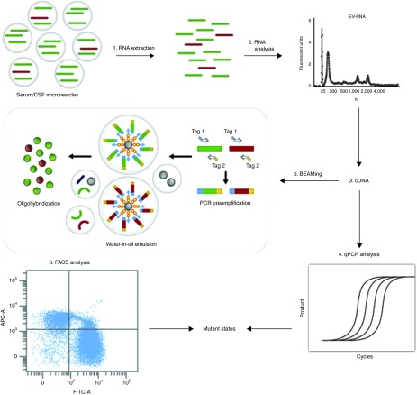
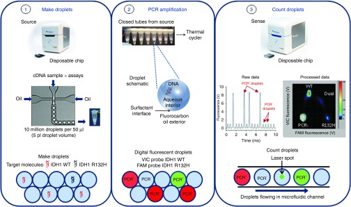
Development of biofluid-based molecular diagnostic tests for cancer is an important step towards tumor characterization and real-time monitoring in a minimally invasive fashion. Extracellular vesicles (EVs) are released from tumor cells into body fluids and can provide a powerful platform for tumor biomarkers because they carry tumor proteins and nucleic acids. Detecting rare point mutations in the background of wild-type sequences in biofluids such as blood and cerebrospinal fluid (CSF) remains a major challenge. Techniques such as BEAMing (beads, emulsion, amplification, magnetics) PCR and droplet digital PCR (ddPCR) are substantially more sensitive than many other assays for mutant sequence detection. Here, we describe a novel approach that combines biofluid EV RNA and BEAMing RT-PCR (EV-BEAMing), as well droplet digital PCR to interrogate mutations from glioma tumors. EVs from CSF of patients with glioma were shown to contain mutant IDH1 transcripts, and we were able to reliably detect and quantify mutant and wild-type IDH1 RNA transcripts in CSF of patients with gliomas. EV-BEAMing and EV-ddPCR represent a valuable new strategy for cancer diagnostics, which can be applied to a variety of biofluids and neoplasms.Molecular Therapy-Nucleic Acids (2013) 2, e109; doi:10.1038/mtna.2013.28; published online 23 July 2013.


Figures

Figure 1
Figure 1
Overview of EV-BEAMing. EVs from serum or CSF samples were pelleted at 100,000g for 80 minutes and processed as follows: (1) RNA was extracted and (2) analyzed for total yield and quality using the Bioanalyzer (Agilent). (3) RNA was then reverse transcribed into cDNA and 1/2 of the sample was used to determine the IDH1 cDNA copy number inside EVs by (4) qPCR analysis. (5) The remaining sample was preamplified (14 cycles) and used as input for BEAMing PCR. The resulting DNA-coated beads were interrogated with sequence-specific fluorescent probes to produce beads with wild-type (green) and mutant (red) profiles. (6) The percentage of beads with mutant DNA was determined by FACS and used in conjunction with the qPCR data to determine the minimum number of copies present to allow reliable detection of the mutant message.
Figure 2
Figure 2
Overview of droplet digital PCR. The RainDrop ddPCR workflow consisted of three steps: (1) PCR reaction mixtures for each sample were pipetted into one of eight wells on the Source microfluidic chip, which was next placed above empty PCR tubes in the RainDrop Source. Pressure-driven air flow forced the aqueous mixture into the chip along with a surfactant-containing fluorocarbon oil to generate five picoliter droplets, and then deposited automatically into the PCR tube strip. (2) The PCR tube strip was placed into a standard thermal cycler for endpoint PCR amplification, with single-target-molecule-containing droplets resulting in specific probe hydrolysis (PCR+) and bright fluorescence and the majority of droplets, containing no target molecule, resulting in only background probe fluorescence (PCR). (3) Each droplet's fluorescence was detected using a Sense microfluidic chip. The fluorescence was detected and processed into a two-dimensional scatter plot display, custom software was used to draw appropriate gates for each droplet endpoint cluster, and the number of droplets within each gate was counted.
Figure 3
Figure 3
Extracellular particle analysis using EM, cryoTEM and Nanosight. Cerebrospinal fluid (CSF), plasma and serum samples were collected upon or before the opening of the dura mater, respectively, processed as described in methods and stored at −80 °C until further processing. (a) CryoTEM of CSF EVs (i, ii), plasma EVs (iii, iv), and regular EM of CSF EVs (v, vi). Scale bars = 100 nm. (b) CSF (n = 3) and serum samples (n = 3) were diluted 1:50 and 1:3,000 respectively in PBS, and size and concentration of particles were assessed using nanoparticle tracking analysis (NanoSight; ±SD).
Figure 4
Figure 4
Correlation of samples characteristics with the detection of the mutant IDH1. (a) BEAMing FACS plot of one representative mutant positive samples indicating the mutant (Q1) and wild-type populations (Q4). (b) qPCR analysis of the IDH1 mRNA copy number in CSF (n = 14) and serum (n = 10) samples from all GBM patients analyzed that are wild-type or mutant for the IDH1 gene (±SEM). (c) Tumor volumes (left panel) and IDH1 mRNA copy numbers (right panel) in 1 ml of CSF from the mutant IDH1 glioma samples analyzed (n = 8), corresponding to detected or non detected, in mRNA as determined by EV-BEAMing assay and ddPCR.

References

    1. Lecomte T, Ceze N, Dorval E, Laurent-Puig P. Circulating free tumor DNA and colorectal cancer. Gastroenterol Clin Biol. 2010;34:662–681. - PubMed
    1. Schwarzenbach H, Hoon DS, Pantel K. Cell-free nucleic acids as biomarkers in cancer patients. Nat Rev Cancer. 2011;11:426–437. - PubMed
    1. Balaj L, Lessard R, Dai L, Cho YJ, Pomeroy SL, Breakefield XO, et al. Tumour microvesicles contain retrotransposon elements and amplified oncogene sequences. Nat Commun. 2011;2:180. - PMC - PubMed
    1. Skog J, Würdinger T, van Rijn S, Meijer DH, Gainche L, Sena-Esteves M, et al. Glioblastoma microvesicles transport RNA and proteins that promote tumour growth and provide diagnostic biomarkers. Nat Cell Biol. 2008;10:1470–1476. - PMC - PubMed
    1. Noerholm M, Balaj L, Limperg T, Salehi A, Zhu LD, Hochberg FH, et al. RNA expression patterns in serum microvesicles from patients with glioblastoma multiforme and controls. BMC Cancer. 2012;12:22. - PMC - PubMed