Review
. 2013 Sep;8(4):888-99.
doi: 10.1007/s11481-013-9489-x. Epub 2013 Jul 25.

Immune-mediated mechanisms in the pathoprogression of amyotrophic lateral sclerosis

Affiliations
Review

Immune-mediated mechanisms in the pathoprogression of amyotrophic lateral sclerosis

Weihua Zhao et al. J Neuroimmune Pharmacol. 2013 Sep.

Abstract

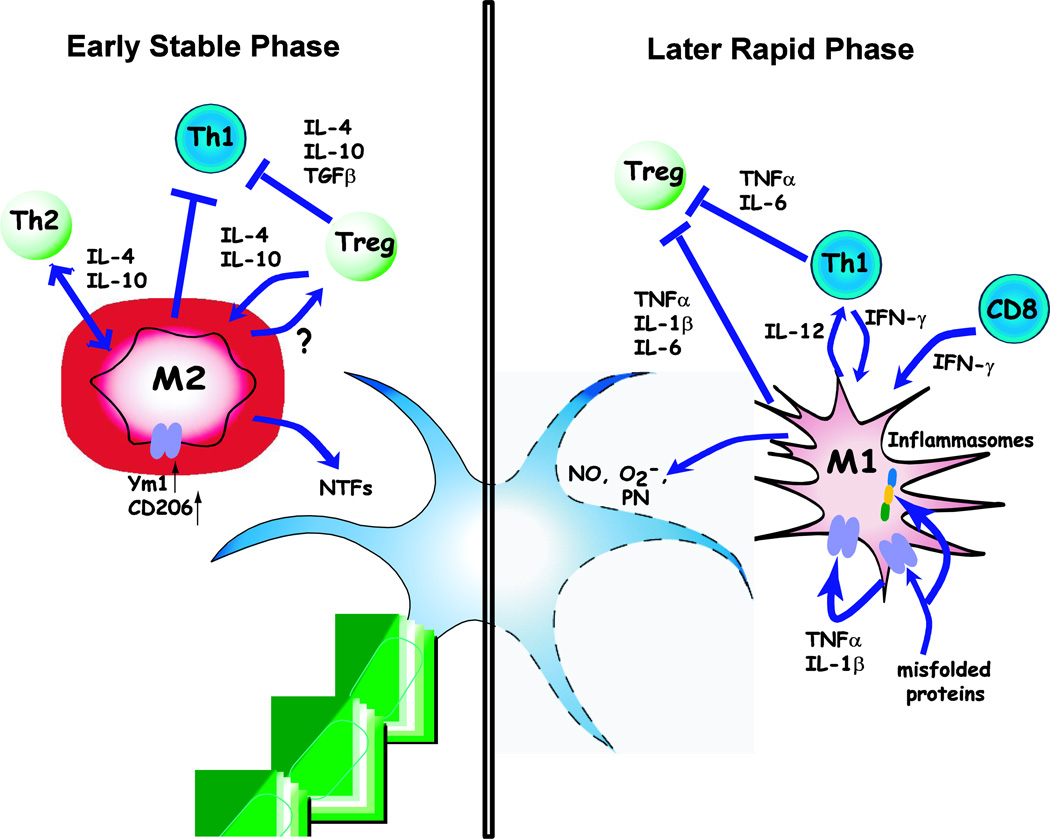
Amyotrophic lateral sclerosis (ALS) is a devastating neurodegenerative disease with selective loss of upper and lower motor neurons. At sites of motor neuron injury, neuroinflammation is a prominent pathological finding and is characterized by microglial activation, astrogliosis, and infiltration of monocytes and T-cells. Both innate and adaptive immune responses actively influence disease progression in animal models and in ALS patients, and promote neuroprotection or neurotoxicity at different stages of disease. The early immune reaction to signals from injured motor neurons is to rescue and repair damaged tissue. As disease accelerates, a shift occurs from beneficial immune responses (involving M2 microglia and regulatory T-cells) to deleterious immune responses (involving M1 microglia and Th1 cells). In this review, we underscore the importance of immune-mediated mechanisms in the pathogenesis of ALS and discuss the alterations and distinct phenotypes of immune cells at the different stages of disease. The better we understand the dynamic changes that occur within the immune system over the course of disease, the better we will be able to develop effective therapeutic regimens in ALS.


Conflict of interest statement

The authors declare that they have no conflict of interest.

Figures

Figure 1
Figure 1
The neuroprotective roles of microglia and astrocytes at early stages of disease. Abnormalities in motor neurons may initiate ALS disease and release repair signals (probably CD200 and fractalkine), which promote an alternatively activated (M2) microglial phenotype. M2 microglia are able to secrete high levels of neurotrophins, such as IGF-1, progranulin, and other neurotrphic factors (NTFs), thereby exerting a neuroprotective function. M2 microglia also release anti-inflammatory cytokines, including IL-1R antagonists and progranulin, to block proinflammatory responses. Astrocytes also participate in the neuroprotective process with secretion of NTFs and uptake of excess glutamate from synaptic clefts. In addition, astrocytes enhance the antioxidant capacity of neurons by releasing the glutathione precursor (CysGly) which is taken up by motor neurons for the synthesis of glutathione. Thus, at early stages, both M2 microglia and astrocytes are involved in sustaining motor neuron health.
Figure 2
Figure 2
The neurotoxic roles of microglia and astrocytes at rapidly progressing stages of disease. As disease progresses, “danger signals” released from motor neurons [possibly mSOD1 or oxidized SOD1 (oxSOD1) and ATP) induce M1 activation of microglia through CD14/TLRs, scavenger receptors (SR), and purinergic P2 receptors. These misfolded proteins may also be endocytosed into microglia to activate inflammasomes. The result of this signaling is a transformation of microglia from an M2 to an M1 phenotype, which produces proinflammatory cytokines (TNF-α and IL-1β, etc), and promotes neurotoxicity by releasing free radicals, including nitric oxide (NO), and superoxide (O2.−). NO and O2.− form the more potent toxin peroxynitrite, which sensitizes AMPA/kainate receptors and increases motor neuron vulnerability to glutamate toxicity. M1 microglia also promote astrocyte activation through ROS and pro-inflammatory cytokines. Activated astrocytes acquire deleterious inflammatory phenotypes with release of ROS, pro-inflammatory cytokines, which, in turn, induce further microglial activation. Thus, ROS production, insufficient NTFs, and impaired glutamate clearance due to down-regulation of GLT1/EAAT2 transporter are the major neurotoxic components of activated astrocytes. Additional unidentified neurotoxic factors released from astrocytes have also been implicated. In addition, M1 microglia, activated astrocytes, and motor neurons produce CCL2, which attracts peripheral M1 monocytes/macrophages into the CNS, and further exacerbates motor neuron degeneration.
Figure 3
Figure 3
In ALS mice, the disease progression curve can be divided into two stages. The early stable phase is associated with beneficial responses of M2 microglia and T regulatory/Th2 cells, while the later rapidly progressing stage is associated with injurious M1 microglia and Th1 responses.
Figure 4
Figure 4
Microglia and T-cell dialogues at different stages of disease. Tregs and Th2 cells predominate at early stable stages of disease. They release anti-inflammatory cytokines, such as IL-4, IL-10, and TGF-β. IL-4 and IL-10 promote an M2 phenotype, characterized by enhanced neurotrophic factors (NTFs) and up-regulation of Ym1 and CD206. M2 microglia in turn promote Treg and Th2 differentiation. In addition, Tregs and M2 microglia also suppress proliferation and cytotoxic function of Th1 cells. Overall, the immune responses at early stages favor neuroprotection through NTFs released from M2 microglia. As disease progression accelerates, the released misfolded proteins and self-propagation give rise to more M1 microglia. The interaction between Th1 and M1 further enhances pro-inflammatory responses, including the release of TNF-α, IL-6, and IL-1β, and downregulated Treg suppressive functions; the beneficial effects of M2 and Tregs/Th2 are lost and the detrimental effects of the Th1 and M1 cells become predominant, enhancing neurodegeneration.

References

    1. Alexianu ME, Kozovska M, Appel SH. Immune reactivity in a mouse model of familial ALS correlates with disease progression. Neurology. 2001;57:1282–1289. - PubMed
    1. Appel SH, Beers DR, Henkel JS. T cell-microglial dialogue in Parkinson's disease and amyotrophic lateral sclerosis: are we listening? Trends Immunol. 2010;31:7–17. - PMC - PubMed
    1. Baloh RH. TDP-43: the relationship between protein aggregation and neurodegeneration in amyotrophic lateral sclerosis and frontotemporal lobar degeneration. FEBS J. 2011;278:3539–3549. - PMC - PubMed
    1. Banerjee R, Mosley RL, Reynolds AD, Dhar A, Jackson-Lewis V, Gordon PH, Przedborski S, Gendelman HE. Adaptive immune neuroprotection in G93A-SOD1 amyotrophic lateral sclerosis mice. PLoS One. 2008;3:e2740. - PMC - PubMed
    1. Barrette B, Hébert MA, Filali M, Lafortune K, Vallières N, Gowing G, Julien JP, Lacroix S. Requirement of myeloid cells for axon regeneration. J Neurosci. 2008;28:9363–9376. - PMC - PubMed

Publication types

MeSH terms

Substances