Comparative Study
. 2013 Dec 15;521(18):4339-55.
doi: 10.1002/cne.23430.

Comparative survey of the topographical distribution of signature molecular lesions in major neurodegenerative diseases

Affiliations
Comparative Study

Comparative survey of the topographical distribution of signature molecular lesions in major neurodegenerative diseases

Steven E Arnold et al. J Comp Neurol. .

Abstract

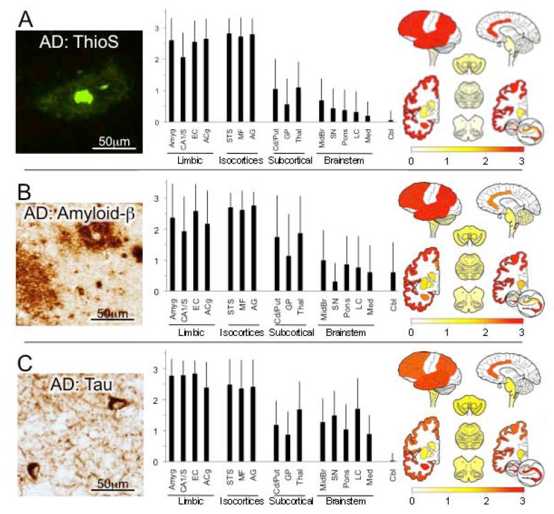
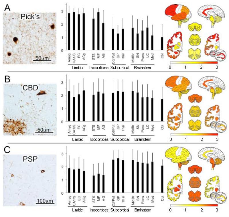
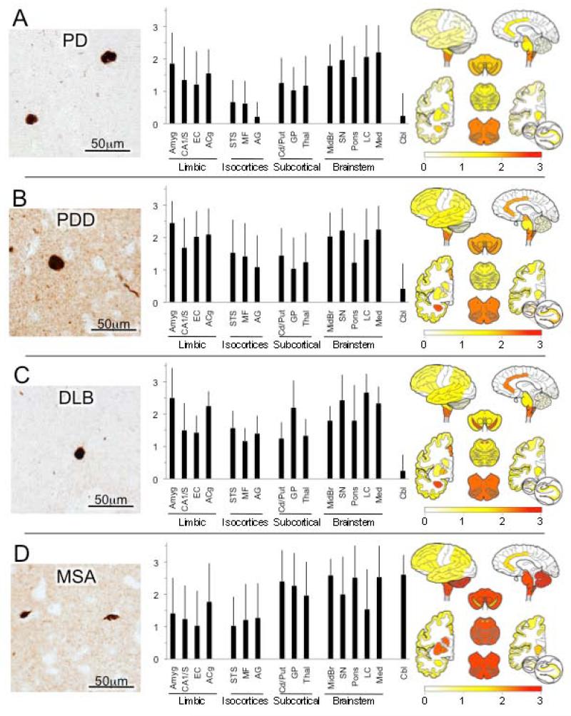
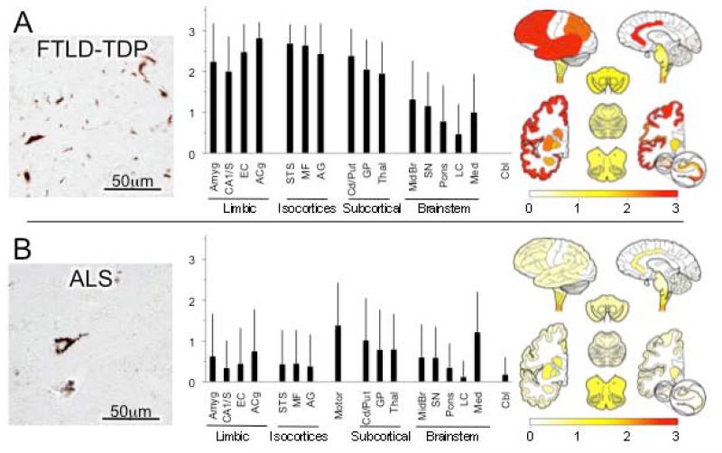
An understanding of the anatomic distributions of major neurodegenerative disease lesions is important to appreciate the differential clinical profiles of these disorders and to serve as neuropathological standards for emerging molecular neuroimaging methods. To address these issues, here we present a comparative survey of the topographical distribution of the defining molecular neuropathological lesions among 10 neurodegenerative diseases from a large and uniformly assessed brain collection. Ratings of pathological severity in 16 brain regions from 671 cases with diverse neurodegenerative diseases are summarized and analyzed. These include: 1) amyloid-β and tau lesions in Alzheimer's disease; 2) tau lesions in three other tauopathies including Pick's disease, progressive supranuclear palsy and corticobasal degeneration; 3) α-synuclein inclusion ratings in four synucleinopathies including Parkinson's disease, Parkinson's disease with dementia, dementia with Lewy bodies, and multiple system atrophy; and 4) TDP-43 lesions in two TDP-43 proteinopathies, including frontotemporal lobar degeneration associated with TDP-43 and amyotrophic lateral sclerosis. The data presented graphically and topographically confirm and extend previous pathological anatomic descriptions and statistical comparisons highlight the lesion distributions that either overlap or distinguish the diseases in each molecular disease category.

Keywords: Alzheimer's disease; Parkinson's disease; Parkinson's disease with dementia; Pick's disease; TDP; TDP-43; Tau α-synuclein; amyloid-β; amyotrophic lateral sclerosis; corticobasal degeneration; dementia with Lewy bodies; frontotemporal lobar degeneration; multiple system atrophy; progressive supranuclear palsy.


Figures

Figure 1
Figure 1
Alzheimer’s disease (AD). (A) Thioflavin S (ThioS)-stained amyloid plaques in mid-frontal gyrus. Photomicrograph shows classical “dense core” plaque with intensely fluorescent amyloid core surrounded by more diffuse amyloid halo. Bar graph depicts average (with standard deviations) ThioS plaque ratings in each of the 16 regions of interest, grouped into neuroanatomic categories (limbic cortices, association isocortices, subcortical nuclei, brainstem and cerebellum). At right, these ratings were projected onto brain mapping template with the rating values indicated in the color bar gradient. (B) Amyloid-β immunohistochemistry. Photomicrograph from mid-frontal gyrus cortex shows major types of plaques - dense cored (upper right) and diffuse (lower left) as well as other scattered amyloid-β deposits. Overall, amyloid-β immunohistochemistry identifies plaque pathology more sensitively than ThioS, as evident in the relatively higher ratings in subcortical nuclei, brainstem and cerebellum. (C) Tau immunohistochemistry. Photomicrograph of angular gyrus shows two neurofibrillary tangles as well as reticular and thread-like neuritic pathology. In graph and map, note very high and consistent tau ratings in limbic cortices but also widespread but more variable involvement throughout brain, including rarely the cerebellum. Abbreviations in graph from left to right: Amyg = amygdala, CA1/S = CA1 and subiculum subfields of hippocampal formation, EC = entorhinal cortex, ACg = anterior cingulate gyrus, STS = superior temporal sulcus including portions of superior and middle temporal gyri, MF = mid-frontal gyrus, AG = angular gyrus, Cd/Put = caudate and putamen, GP = globus pallidus, Thal = thalamus and subthalamus, Midbr = midbrain, SN = substantia nigra, Pons = pons, LC = locus ceruleus, Med = medulla, Cbl = cerebellum.
Figure 2
Figure 2
Tauopathies.* (A) Pick’s disease. Photomicrograph of shows 3 Pick bodies as well as diffuse neuritic pathology in background. As shown in the graph and map, tau pathology is greatest in limbic and isocortical association areas, but can also be found throughout the brain. (B) Corticobasal degeneration (CBD). Photomicrograph shows tau-immunoreactive tangle-line neuronal inclusion (upper right) and astrocytic “plaque” (lower left) as well as scattered immunoreactive neuritic/glial processes. As depicted in the graph and map, tau pathology is widely distributed. (C) Progressive supranuclear palsy (PSP). Global ratings were assigned based on abnormal tau inclusions in glia and neurons as well as glial and neuronal processes. In contrast to Pick’s disease and AD, graphs portray the predominant subcortical nuclei and hindbrain pathology of PSP. *All photomicrographs are from mid-frontal gyrus.
Figure 3
Figure 3
α-Synucleinopathies.* (A) Parkinson’s disease (PD). While predominant in brainstem, Lewy pathology is often found in subcortical nuclei and cortex, especially limbic cortex. Photomicrograph shows Lewy bodies in mid-frontal gyrus. (B) Parkinson’s Disease Dementia (PDD). α-Synuclein pathology in the form of intraneuronal Lewy bodies and dystrophic Lewy neurites (photomicrograph shows mid-frontal cortex) is greater and more widely disseminated, especially in cerebral cortex in PDD. (C) Dementia with Lewy bodies (DLB) shows virtually identical pathology and distribution pattern to PDD. (D) Multiple system atrophy (MSA). The pathological α-synuclein lesions that predominate in MSA are oligodendrocyte glial cell inclusions (GCIs, photomicrograph) that distinguish MSA from PD, PDD and DLB. GCIs as well as some Lewy-like neuronal pathology are widespread in MSA but predominate in hindbrain and subcortical nuclei. *All photomicrographs are from mid-frontal gyrus.
Figure 4
Figure 4
TDP-43 proteinopathies.* (A) Frontotemporal lobar degeneration associated with TDP-43 (FTDL-TDP). Neuronal and neuritic pathological TDP-43 inclusions of various morphologies (photomicrograph) severely affect limbic and isocortical association cortices as well as subcortical nuclei, more so than brain stem. TDP-43 lesions were not observed in cerebellum in any case. (B) Amyotrophic lateral sclerosis (ALS). Photomicrograph shows neuron with diffuse perikaryal TDP-43 immunoreactivity. Graph and map depict widespread but generally mild lesion ratings throughout the brain in ALS with exception of higher ratings motor cortex and lower brainstem. In addition, cervical spinal cord was commonly examined in ALS cases and showed high ratings (data not shown). *Both photomicrographs are from mid-frontal gyrus.

References

    1. Aarsland D, Andersen K, Larsen JP, Lolk A, Kragh-Sorensen P. Prevalence and characteristics of dementia in Parkinson disease: an 8-year prospective study. Arch Neurol. 2003;60(3):387–392. - PubMed
    1. Aarsland D, Zaccai J, Brayne C. A systematic review of prevalence studies of dementia in Parkinson’s disease. Mov Disord. 2005;20(10):1255–1263. - PubMed
    1. Alafuzoff I, Arzberger T, Al-Sarraj S, Bodi I, Bogdanovic N, Braak H, Bugiani O, Del-Tredici K, Ferrer I, Gelpi E, Giaccone G, Graeber MB, Ince P, Kamphorst W, King A, Korkolopoulou P, Kovacs GG, Larionov S, Meyronet D, Monoranu C, Parchi P, Patsouris E, Roggendorf W, Seilhean D, Tagliavini F, Stadelmann C, Streichenberger N, Thal DR, Wharton SB, Kretzschmar H. Staging of neurofibrillary pathology in Alzheimer’s disease: a study of the BrainNet Europe Consortium. Brain Pathol. 2008;18(4):484–496. - PMC - PubMed
    1. Alzheimer’s Association Alzheimer’s disease facts and figures. Alzheimer’s & dementia: the journal of the Alzheimer’s Association. 2013;9(2):208–245. - PubMed
    1. Armstrong MJ, Litvan I, Lang AE, Bak TH, Bhatia KP, Borroni B, Boxer AL, Dickson DW, Grossman M, Hallett M, Josephs KA, Kertesz A, Lee SE, Miller BL, Reich SG, Riley DE, Tolosa E, Troster AI, Vidailhet M, Weiner WJ. Criteria for the diagnosis of corticobasal degeneration. Neurology. 2013;80(5):496–503. - PMC - PubMed

Publication types