. 2013 Sep;19(9):1238-52.
doi: 10.1261/rna.039255.113. Epub 2013 Jul 23.

The double-stranded RNA binding domain of human Dicer functions as a nuclear localization signal

Affiliations

The double-stranded RNA binding domain of human Dicer functions as a nuclear localization signal

Michael Doyle et al. RNA. 2013 Sep.

Abstract

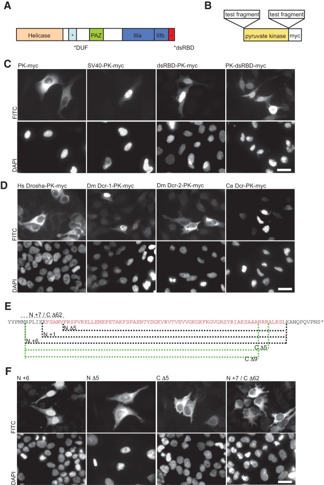
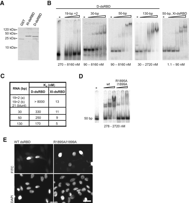
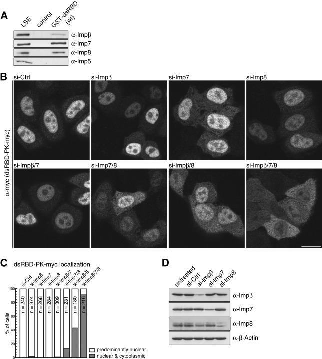
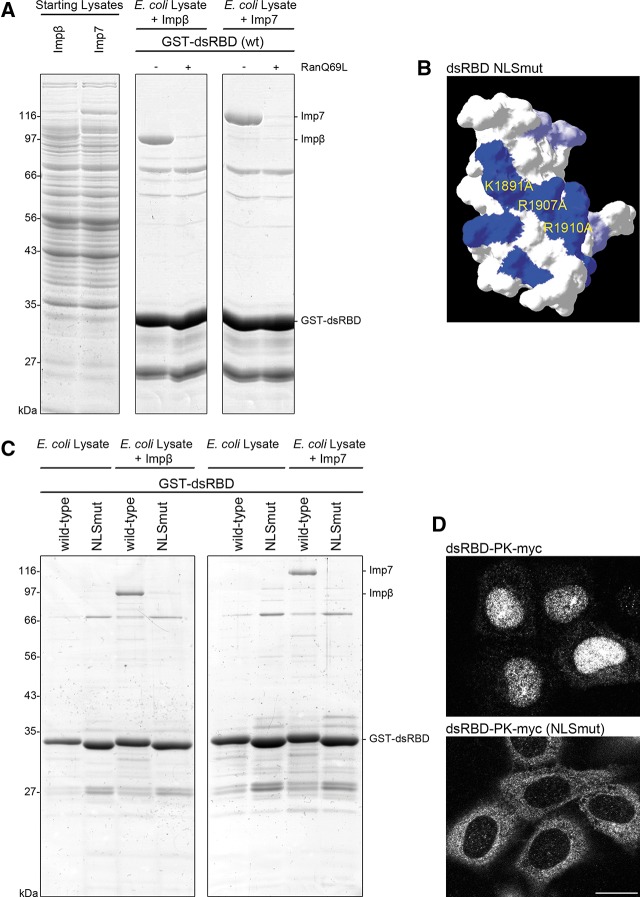
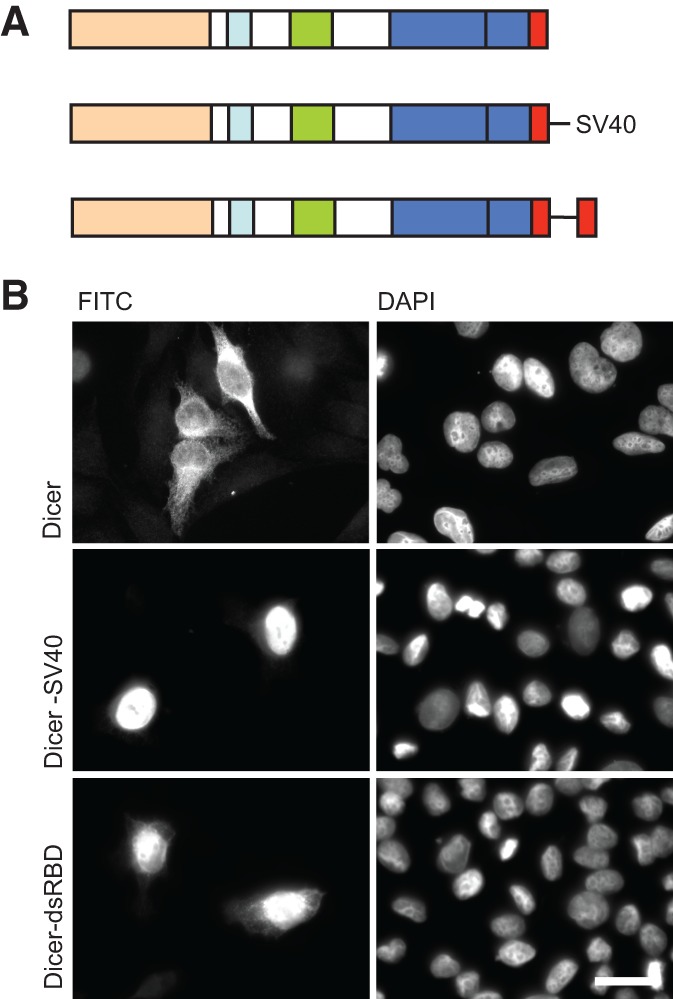
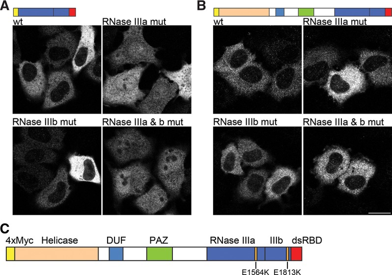
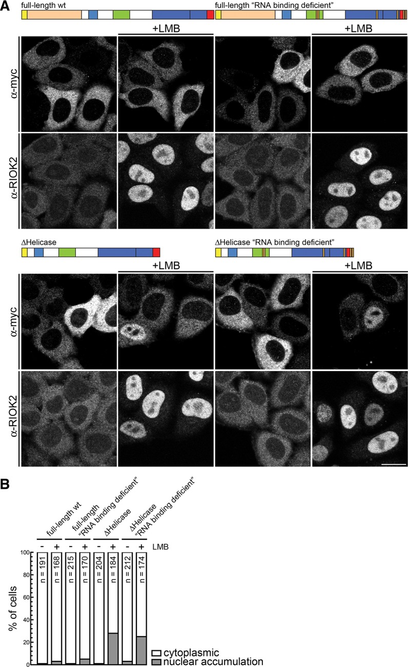
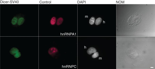
Dicer is a key player in microRNA (miRNA) and RNA interference (RNAi) pathways, processing miRNA precursors and double-stranded RNA into ∼21-nt-long products ultimately triggering sequence-dependent gene silencing. Although processing of substrates in vertebrate cells occurs in the cytoplasm, there is growing evidence suggesting Dicer is also present and functional in the nucleus. To address this possibility, we searched for a nuclear localization signal (NLS) in human Dicer and identified its C-terminal double-stranded RNA binding domain (dsRBD) as harboring NLS activity. We show that the dsRBD-NLS can mediate nuclear import of a reporter protein via interaction with importins β, 7, and 8. In the context of full-length Dicer, the dsRBD-NLS is masked. However, duplication of the dsRBD localizes the full-length protein to the nucleus. Furthermore, deletion of the N-terminal helicase domain results in partial accumulation of Dicer in the nucleus upon leptomycin B treatment, indicating that CRM1 contributes to nuclear export of Dicer. Finally, we demonstrate that human Dicer has the ability to shuttle between the nucleus and the cytoplasm. We conclude that Dicer is a shuttling protein whose steady-state localization is cytoplasmic.

Keywords: Dicer; NLS; RNAi; dsRBD; helicase.


Figures

FIGURE 1.
FIGURE 1.
The dsRBD of human Dicer functions as a nuclear localization signal (NLS). (A) Schematic representation of human Dicer with individual protein domains indicated by different colors. (B) The pyruvate kinase (PK-myc) fusion constructs used to assess NLS activity. (C) In transfected HeLa cells, PK-myc localizes to the cytoplasm but can be driven to the nucleus by an SV40 NLS (SV40-PK-myc). dsRBD-PK-myc and PK-dsRBD-myc show strong nuclear accumulation. Staining with DAPI is shown in the lower panel. (D) dsRBDs from various proteins were tested for their ability to function as a NLS in the PK reporter assay as indicated. The dsRBDs from human Drosha (Hs Drosha-PK-myc) and Drosophila Dcr-2 (Dm Dcr-2-PK-myc) were found not to function as a NLS, whereas those of Drosophila Dcr-1 (Dm Dcr-1-PK-myc) and C. elegans Dcr (Ce Dcr-PK-myc) functioned as a NLS. Bar, 20 µm. (E) Sequence of Dicer’s dsRBD (amino acids 1845 to 1912, in red) and its flanking amino acids (in black). The indicated deletions made at either the N or C terminus of the dsRBD are shown by dashed lines. The protein fragments of each dsRBD reporter tested are as follows: N Δ5, 1850 to 1912; N +1, 1844 to 1912; N +6, 1839 to 1912; C Δ5, 1839 to 1907; C Δ9, 1839 to 1903. N +7/C Δ62, including only residues 1837 to 1849, served as an additional control to test if residues spanning gaps between other deletion constructs can function as a NLS. (F) In transfected HeLa cells, Dicer dsRBD N +6 and N +1 (data not shown for N +1) accumulated in the nucleus. N- or C-terminal deletions (N Δ5, C Δ5, and C Δ9 [data not shown for C Δ9]) localize to the cytoplasm. The additional control N +7/C Δ62 also localizes to the cytoplasm. Staining with DAPI is shown in the lower panel. Representative images are shown from a minimum of three independent experiments. Bar, 20 µm.
FIGURE 2.
FIGURE 2.
RNA-binding potential of Dicer dsRBD is not required for NLS activity. (A) Dicer’s dsRBD (D-dsRBD) and the second dsRBD from the Xenopus laevis RNA-binding protein A (XlrbpA) (Xl-dsRBD) were expressed as GST-fusion proteins and purified from Escherichia coli. (B) To analyze the D-dsRBD RNA-binding properties, native gel mobility shift assays were performed three or more times with increasing amounts of recombinant D-dsRBD and substrates of differing length as indicated. D-dsRBD bound 30-, 50-, and 130-bp-long dsRNAs, but only little binding of the shorter 21- or 19-bp substrates was observed at protein concentrations tested. Two independent sequences for the 19-bp substrates containing 2-nt 3′-overhangs, 19 + 2 (a) and 19 + 2 (b), were tested. (C) Apparent binding affinities for the different substrates were measured for D-dsRBD and the Xl-dsRBD using the representative experiments shown in panel B. D-dsRBD bound longer dsRNAs with an affinity ∼30 times lower compared to the Xl-dsRBD. (D) Mutational analysis of D-dsRBD was performed (also see Supplemental Fig. S2) to identify residues important for RNA-binding. Introduction of the double point mutation (R1895A/I1899A) severely abrogated RNA-binding. (E) The NLS activity of the double point mutant (R1895A/I1899A) was tested in the PK-myc reporter assay and compared to the wild-type (WT) domain. Both fusion proteins efficiently accumulated in the nucleus. Staining with DAPI is shown in the lower panel. Representative images are shown from a minimum of three independent experiments. Bar, 20 µm.
FIGURE 3.
FIGURE 3.
The dsRBD of human Dicer binds the nuclear transport receptors Impβ, Imp7, and Imp8. (A) Recombinant, purified GST-dsRBD (wt) was immobilized on glutathione (GSH)-sepharose beads and incubated with low-salt HeLa cell extract (LSE). Bound proteins were eluted and separated by SDS-PAGE, followed by immunoblotting using antibodies directed to human Impβ, Imp7, Imp8, and Imp5. Control, GSH-sepharose beads alone. (B) Simultaneous depletion of Impβ, Imp7, and Imp8 results in a cytoplasmic accumulation of dsRBD-PK-myc. Tetracycline-inducible dsRBD-PK-myc reporter cells were transfected with indicated siRNAs. Sixty hours after transfection, PK-dsRBD-myc expression was induced for 12 h. Cells were then fixed, followed by immunofluorescence (IF) using an α-myc antibody. Bar, 20 μm. (C) Quantification of the experiment shown in B. Cells displaying predominantly nuclear or nuclear and cytoplasmic localization of dsRBD-PK-myc were counted. (N) Number of counted cells. (D) HeLa cells were transfected with siRNAs targeting different importins. Seventy-two hours post-transfection, cells were harvested, and the protein levels were analyzed by Western blotting. β-actin served as a loading control.
FIGURE 4.
FIGURE 4.
Dicer’s dsRBD contains a basic region critical for NLS activity. (A) Recombinant GST-dsRBD (wt) was immobilized on GSH-sepharose beads and mixed with the indicated purified factors (Impβ, Imp7) added to E. coli lysate. Bound proteins were eluted and separated by SDS-PAGE followed by Coomassie staining. (B) Representation of the basic patch in Dicer’s dsRBD (blue, with positions of mutated residues indicated). Mutation of its amino acid residues to alanines destroyed the dsRBD NLS activity. Mutation of two additional basic residues, K1887 and K1889, to alanines had no appreciable effect on nuclear localization of the dsRBD-PK reporter (data not shown). (C) Binding of Impβ and Imp7 to Dicer’s dsRBD is dependent on the integrity of the basic patch. The experiment was performed as described in A, comparing Impβ and Imp7 recruitment to the wt and NLSmut dsRBD. (D) HeLa cells were transiently transfected with dsRBD-PK-myc or dsRBD-PK-myc NLSmut (K1891A/R1907A/R1910A). Twenty-four hours post-transfection, cells were fixed and immunofluorescence (IF) was performed with an α-myc antibody. Representative images are shown from a minimum of three independent experiments. Bar, 20 μm.
FIGURE 5.
FIGURE 5.
Dicer dsRBD is occluded in the full-length protein. (A) Schematic representation of human Dicer and constructs which have been tested for localization. (B) Transfection of HeLa cells reveals that nuclear localization of full-length Dicer (Dicer) can be achieved either by the addition of an SV40 NLS at the C terminus (Dicer-SV40) or by duplicating the dsRBD (Dicer-dsRBD). Representative images are shown from a minimum of three independent experiments. Bar, 20 µm.
FIGURE 6.
FIGURE 6.
RNA-binding influences Dicer localization. (A) Transfection of HeLa cells with the C-terminal fragment of Dicer (RNase III domains plus dsRBD). The wild-type (wt) construct fails to localize to the nucleus. When E1564K is introduced into RNase IIIa catalytic core (RNase IIIa mut), nuclear accumulation can be observed. A similar mutation (E1813K) in the catalytic core of RNase IIIb (RNase IIIb mut) also leads to partial localization in the nucleus, although the effect is much weaker compared to RNase IIIa mut. The double mutation E1564K/E1813K showed strong nuclear accumulation. (B) Transfection of HeLa cells with full-length Dicer constructs. Wild-type (wt) Dicer localizes to the cytoplasm. Introduction of mutations E1564K or E1813K into the RNase III domains either individually (RNase IIIa mut and RNase IIIb mut) or together (RNase IIIa & b mut) showed cytoplasmic staining similar to wt protein. Representative images are shown from a minimum of three independent experiments. Bar, 20 µm. (C) Schematic representation of human Dicer with individual protein domains indicated by different colors and the position of each RNase III mutation shown.
FIGURE 7.
FIGURE 7.
The helicase domain plays a central role in the cytoplasmic retention of Dicer. (A) HeLa cells were transfected with the indicated constructs. Twenty-four hours post transfection, cells were treated with 20 nM LMB or solvent (ethanol) for 10 h and fixed, followed by IF using an α-myc antibody. IF against RIOK2 served as a positive control for the LMB treatment. Bar, 20 μm. (B) Quantification of the experiment shown in A. Cells displaying either cytoplasmic localization or nuclear accumulation of the constructs were counted. (N) Number of counted cells.
FIGURE 8.
FIGURE 8.
Dicer has the potential to shuttle. Heterokaryon analysis of Dicer-SV40. HeLa cells were cotransfected with plasmids expressing Dicer-SV40 and either hnRNPA1 (positive shuttling control; top panel) or hnRNPC (negative shuttling control; bottom panel). The transfected cells were fused with nontransfected mouse NIH 3T3 cells. Dicer-SV40 was detected in both HeLa and NIH 3T3 nuclei (top and bottom panels) as was hnRNPA1 (top panel). hnRNPC was only detected in the HeLa nuclei (bottom panel). Nuclei were distinguished by DAPI staining and are indicated. (h) Human HeLa cells, (m) mouse NIH 3T3 cells. Bar, 5 µm.

Similar articles

Cited by

References

    1. Ando Y, Tomaru Y, Morinaga A, Burroughs AM, Kawaji H, Kubosaki A, Kimura R, Tagata M, Ino Y, Hirano H, et al. 2011. Nuclear pore complex protein mediated nuclear localization of dicer protein in human cells. PLoS One 6: e23385. - PMC - PubMed
    1. Barraud P, Emmerth S, Shimada Y, Hotz HR, Allain FH, Buhler M 2011. An extended dsRBD with a novel zinc-binding motif mediates nuclear retention of fission yeast Dicer. EMBO J 30: 4223–4235 - PMC - PubMed
    1. Benetti R, Gonzalo S, Jaco I, Munoz P, Gonzalez S, Schoeftner S, Murchison E, Andl T, Chen T, Klatt P, et al. 2008. A mammalian microRNA cluster controls DNA methylation and telomere recombination via Rbl2-dependent regulation of DNA methyltransferases. Nat Struct Mol Biol 15: 268–279 - PMC - PubMed
    1. Billy E, Brondani V, Zhang H, Muller U, Filipowicz W 2001. Specific interference with gene expression induced by long, double-stranded RNA in mouse embryonal teratocarcinoma cell lines. Proc Natl Acad Sci 98: 14428–14433 - PMC - PubMed
    1. Blaszczyk J, Gan J, Tropea JE, Court DL, Waugh DS, Ji X 2004. Noncatalytic assembly of ribonuclease III with double-stranded RNA. Structure 12: 457–466 - PubMed

Publication types

MeSH terms

LinkOut - more resources