. 2013 Jul 17:4:185.
doi: 10.3389/fphys.2013.00185. eCollection 2013.

Membrane potential and cancer progression

Affiliations

Membrane potential and cancer progression

Ming Yang et al. Front Physiol. .

Abstract

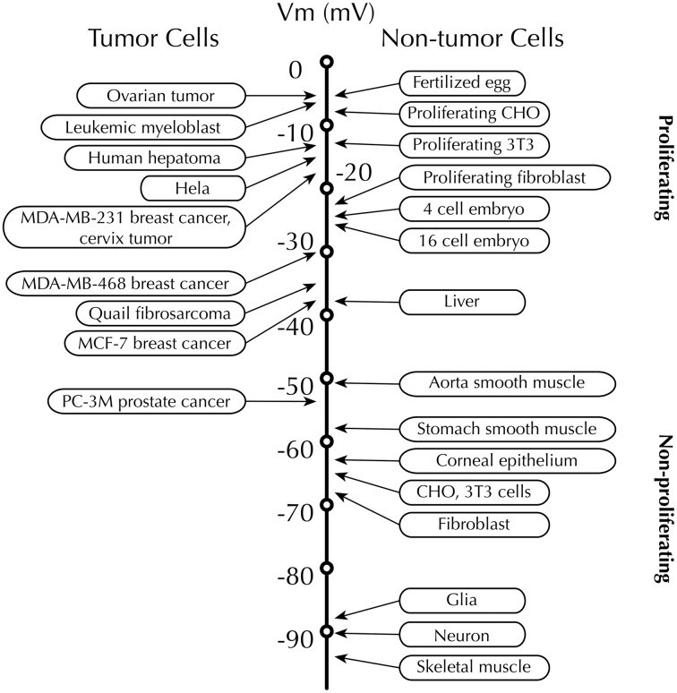
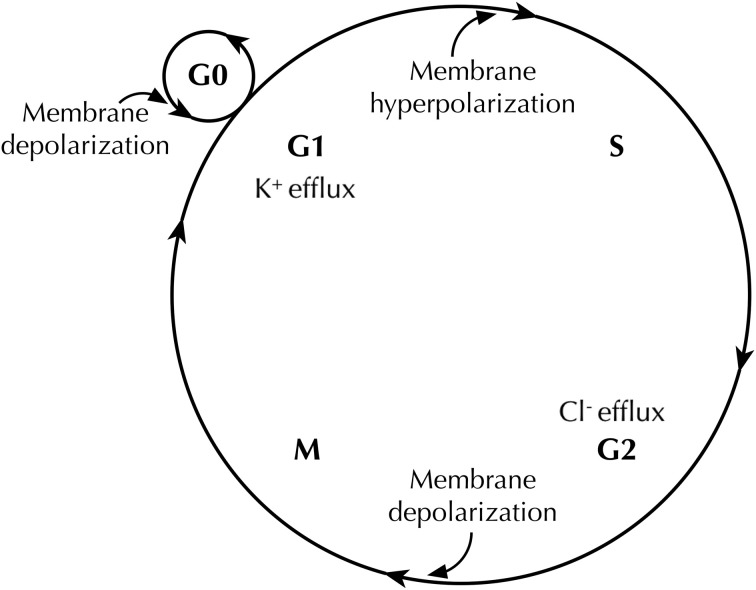
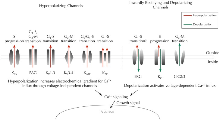
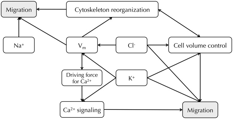
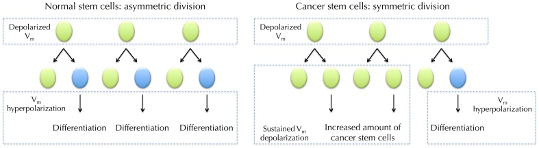
Membrane potential (Vm ), the voltage across the plasma membrane, arises because of the presence of different ion channels/transporters with specific ion selectivity and permeability. Vm is a key biophysical signal in non-excitable cells, modulating important cellular activities, such as proliferation and differentiation. Therefore, the multiplicities of various ion channels/transporters expressed on different cells are finely tuned in order to regulate the Vm . It is well-established that cancer cells possess distinct bioelectrical properties. Notably, electrophysiological analyses in many cancer cell types have revealed a depolarized Vm that favors cell proliferation. Ion channels/transporters control cell volume and migration, and emerging data also suggest that the level of Vm has functional roles in cancer cell migration. In addition, hyperpolarization is necessary for stem cell differentiation. For example, both osteogenesis and adipogenesis are hindered in human mesenchymal stem cells (hMSCs) under depolarizing conditions. Therefore, in the context of cancer, membrane depolarization might be important for the emergence and maintenance of cancer stem cells (CSCs), giving rise to sustained tumor growth. This review aims to provide a broad understanding of the Vm as a bioelectrical signal in cancer cells by examining several key types of ion channels that contribute to its regulation. The mechanisms by which Vm regulates cancer cell proliferation, migration, and differentiation will be discussed. In the long term, Vm might be a valuable clinical marker for tumor detection with prognostic value, and could even be artificially modified in order to inhibit tumor growth and metastasis.

Keywords: cancer; cell cycle; differentiation; ion channel; membrane potential; migration; proliferation; stem cell.


Figures

Figure 1
Figure 1
Membrane potential (Vm) scale. Rapidly proliferating cancer cells possess depolarized Vm, while the Vm of quiescent cells is generally more negative. Proliferative somatic cells are also depolarized, suggesting that Vm is functionally instructive in cell development (Levin, 2007b). Scale adapted from Binggeli and Weinstein (1986), with additional data from Fraser et al. (2005); Mycielska et al. (2005); Yang et al. (2012).
Figure 2
Figure 2
Membrane potential (Vm) changes during the cell cycle. Vm undergoes hyperpolarization at G1/S border, by virtue of K+ efflux through various K+ channels. Before cells enter M phase, increased Cl efflux accompanies Vm depolarization. Quiescent cells at G0 stage show mitotic activities after Vm depolarization (Cone and Cone, 1976).
Figure 3
Figure 3
Key ion channels that regulate Vm and cell cycle progression in cancer. Hyperpolarizing channels (outward IK, red) would increase the driving force for Ca2+ influx through voltage-independent channels, whereas inwardly rectifying K+ channels (predominantly inward IK, green) and chloride channels (outward Cl, green) would depolarize the Vm, thus enabling activation of voltage-dependent Ca2+ influx (Schwab et al., 2012). Time- and domain-dependent Ca2+ signaling is then proposed to activate pathways that promote cell cycle progression and proliferation. Abbreviations: KCa, Ca2+-activated K+ channel; EAG, ether à go-go channel; Kv, voltage-gated K+ channel; KATP, ATP-sensitive K+ channel; K2P, two-pore domain K+ channel; ERG, EAG-related gene K+ channel; Kir, classic inward-rectifier K+ channel; ClC2/3, chloride 2/3 channel.
Figure 4
Figure 4
Relationship between Na+, K+, Cl channels and Vm in cancer cell migration. Vm provides the driving force for Ca2+, and downstream Ca2+ signaling leads to cell migration (Schwab et al., 2012). Vm also regulates cytoskeleton reorganization (Chifflet et al., 2003, 2004). Cl and K+ channels both contribute to Vm regulation and cell volume control (Soroceanu et al., ; Sontheimer, ; Habela et al., ; Schwab et al., 2012). Inhibiting particular Na+, K+, and Cl channels can reduce cancer cell migration (Sontheimer, ; Brackenbury, ; Schwab et al., 2012).
Figure 5
Figure 5
Vm in normal stem cell (SC) differentiation and hypothesized role for Vm in cancer stem cells (CSCs). Depolarized Vm is needed during the maintenance of SCs. SC undergoes asymmetric division where it produces one copy of itself and one progeny that later differentiate into mature cells. The maturation requires Vm hyperpolarization (Sundelacruz et al., 2008). However, CSCs frequently undergo symmetric division, in which one CSC divides into two identical CSC progenies (Wicha et al., 2006). Sustained Vm depolarization may help to maintain the increasing CSCs in an undifferentiated state. Proliferation of CSCs then increases cancer malignancy.

References

    1. Abdul M., Hoosein N. (2002). Expression and activity of potassium ion channels in human prostate cancer. Cancer Lett. 186, 99–105 10.1016/S0304-3835(02)00348-8 - DOI - PubMed
    1. Adams D. S. (2008). A new tool for tissue engineers: ions as regulators of morphogenesis during development and regeneration. Tissue Eng. Part A 14, 1461–1468 10.1089/ten.tea.2008.0080 - DOI - PubMed
    1. Adams D. S., Levin M. (2012). General principles for measuring resting membrane potential and ion concentration using fluorescent bioelectricity reporters. Cold Spring Harb. Protoc. 2012, 385–397 - PMC - PubMed
    1. Adams D. S., Robinson K. R., Fukumoto T., Yuan S., Albertson R. C., Yelick P., et al. (2006). Early, H+-V-ATPase-dependent proton flux is necessary for consistent left-right patterning of non-mammalian vertebrates. Development 133, 1657–1671 10.1242/dev.02341 - DOI - PMC - PubMed
    1. Arcangeli A. (2005). Expression and role of hERG channels in cancer cells. Novartis Found. Symp. 266, 225–232 discussion: 232–234. 10.1002/047002142X.ch17 - DOI - PubMed