. 2013 Jul 25;3(1):56.
doi: 10.1186/2191-219X-3-56.

Post-radioembolization yttrium-90 PET/CT - part 1: diagnostic reporting

Affiliations

Post-radioembolization yttrium-90 PET/CT - part 1: diagnostic reporting

Yung-Hsiang Kao et al. EJNMMI Res. .

Abstract

Background: Yttrium-90 (90Y) positron emission tomography with integrated computed tomography (PET/CT) represents a technological leap from 90Y bremsstrahlung single-photon emission computed tomography with integrated computed tomography (SPECT/CT) by coincidence imaging of low abundance internal pair production. Encouraged by favorable early experiences, we implemented post-radioembolization 90Y PET/CT as an adjunct to 90Y bremsstrahlung SPECT/CT in diagnostic reporting.

Methods: This is a retrospective review of all paired 90Y PET/CT and 90Y bremsstrahlung SPECT/CT scans over a 1-year period. We compared image resolution, ability to confirm technical success, detection of non-target activity, and providing conclusive information about 90Y activity within targeted tumor vascular thrombosis. 90Y resin microspheres were used. 90Y PET/CT was performed on a conventional time-of-flight lutetium-yttrium-oxyorthosilicate scanner with minor modifications to acquisition and reconstruction parameters. Specific findings on 90Y PET/CT were corroborated by 90Y bremsstrahlung SPECT/CT, 99mTc macroaggregated albumin SPECT/CT, follow-up diagnostic imaging or review of clinical records.

Results: Diagnostic reporting recommendations were developed from our collective experience across 44 paired scans. Emphasis on the continuity of care improved overall diagnostic accuracy and reporting confidence of the operator. With proper technique, the presence of background noise did not pose a problem for diagnostic reporting. A counter-intuitive but effective technique of detecting non-target activity is proposed, based on the pattern of activity and its relation to underlying anatomy, instead of its visual intensity. In a sub-analysis of 23 patients with a median follow-up of 5.4 months, 90Y PET/CT consistently outperformed 90Y bremsstrahlung SPECT/CT in all aspects of qualitative analysis, including assessment for non-target activity and tumor vascular thrombosis. Parts of viscera closely adjacent to the liver remain challenging for non-target activity detection, compounded by a tendency for mis-registration.

Conclusions: Adherence to proper diagnostic reporting technique and emphasis on continuity of care are vital to the clinical utility of post-radioembolization 90Y PET/CT. 90Y PET/CT is superior to 90Y bremsstrahlung SPECT/CT for the assessment of target and non-target activity.


Figures

Figure 1
Figure 1
Patient 23. (a) Multifocal HCC seen on catheter-directed CT angiogram of the replaced left hepatic artery, arising from the left gastric artery. Four small tumors are indicated by arrows. (b) Enlarged view of segment IV shows the larger central tumor being flanked by two smaller, closely adjacent tumors separated by approximately 3 mm. (c) Post-radioembolization 90Y PET/CT confirms focal activity within the four small tumors, depicted in high resolution. (d)90Y bremsstrahlung SPECT/CT was barely able to detect subtle activity in the larger (arrow) of the four small tumors, but was unable to confirm any focal activity within the other three small tumors due to its low resolution.
Figure 2
Figure 2
Patient 20. (a) Large hypovascular HCC in the right lobe, seen on contrast-enhanced CT Abdomen. (b) Catheter-directed CT angiogram of the right hepatic artery demonstrates tumor hypovascularity and delineates the target arterial territory. (c)90Y PET/CT depicts, in high resolution, a rim-like activity in the tumor periphery with a large area of relative central photopenia, typical of hypovascular tumors. (d)90Y bremsstrahlung SPECT/CT poorly demonstrates the peripheral activity due to its low image resolution and under-represents the extent of central photopenia.
Figure 3
Figure 3
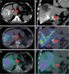
Patient 10. Portal vein tumor thrombosis in a large heterogeneous HCC. (a, b) Catheter-directed CT angiogram of the right hepatic artery demonstrates contrast enhancement within portal vein tumor thrombosis (arrow). (c, d)90Y PET/CT depicts focal 90Y activity within the portal vein tumor thrombus in high resolution. (e, f) The same activity is poorly visualized on 90Y bremsstrahlung SPECT/CT as subtle, ill-defined activity.
Figure 4
Figure 4
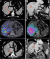
Patient 12. (a, b) Portal vein tumor thrombosis (arrow) in a multifocal HCC seen on portal venous phase of a triphasic CT liver. (c)90Y PET/CT demonstrates absent activity within the portal vein tumor thrombus. (d) The diffuse nature of 90Y bremsstrahlung SPECT/CT is unable to resolve the absence of activity within the portal vein tumor thrombus. (e, f) Four months post-radioembolization, a follow-up triphasic CT liver in the portal venous phase shows significant progression of portal vein tumor thrombosis, validating the 90Y PET/CT findings.
Figure 5
Figure 5
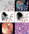
Patient 17.90 Y radioembolization of a recurrent intra-hepatic cholangiocarcinoma. (a) Pre-radioembolization coeliac axis ‘C’ digital subtraction angiography (DSA) shows the common hepatic artery bifurcating into the right hepatic and gastroduodenal arteries ‘GDA’, later branching into the right gastroepiploic artery ‘RGE’. DSA at the conclusion of 90Y radioembolization demonstrated significant vascular stasis and reflux of contrast into the gastroduodenal and right gastroepiploic arteries (images not shown). (b) Coronal view of post-radioembolization 90Y PET/CT and its (c) MIP demonstrates in high resolution, non-target activity along the gastric greater curve ‘G’, antrum ‘A’ and proximal duodenum ‘D’. (d) MIP of 90Y bremsstrahlung SPECT shows concordant non-target activity, but of lower intensity and resolution. By quantification 90Y PET activity, detailed in Part 2 [38], the non-target mean absorbed doses to the gastric greater curve, gastric outlet, and proximal duodenum were approximately 49, 65, and 53 Gy, respectively. Within weeks, the patient developed persistent epigastric pain. (e) Gastroscopy at 3.3 months revealed extensive inflammation along the stomach and proximal duodenum. A Forrest III ulcer was present in the pylorus (large arrow). The ulcer edge (small arrows) was inflamed and indurated. (f) Photomicrograph (H&E stain; ×200) of the gastric biopsy showed edema, congestion, and diffuse lymphoplasmacytic, eosinophilic, and neutrophilic infiltrates. A purple-staining resin microsphere (arrow) surrounded by a halo was seen in the lamina propria. The surrounding gastric glands show flattening of the epithelium, dilatation, and reactive atypia, consistent with severe cellular injury.
Figure 6
Figure 6
Patient 22.90 Y radioembolization of part of a segment III HCC via the terminal branch of the right internal mammary artery (RIMA). (a, b) Pre-therapy 99mTc MAA SPECT/CT demonstrates non-target activity in the right lower anterior chest wall (arrows), shunted from proximal branches of the RIMA. (c, d)90Y PET/CT depicts in high-resolution, non-target activity conforming to the anatomy of the right lower anterior chest wall, concordant with 99mTc MAA SPECT/CT. The slight dissimilarity in chest wall activity biodistribution between 90Y PET/CT versus 99mTc MAA SPECT/CT is due to local arterial flow changes after prophylactic bland embolization of proximal branches of the RIMA (images not shown). (e, f)90Y bremsstrahlung SPECT/CT was unable to detect any non-target activity in the chest wall due to its low image resolution.
Figure 7
Figure 7
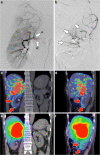
Patient 9.90 Y radioembolization of an organ other than the liver. KIT-negative GIST with bulky metastasis to the right adrenal gland, refractory to tyrosine kinase inhibitors. (a) Pre-radioembolization digital subtraction angiogram (DSA) with catheter tip in the right renal artery ‘R’ demonstrates the origin of the right inferior adrenal artery ‘A’ and the arterial trees of the adrenal tumor and right kidney. The catheter tip was advanced deep into the right inferior adrenal artery for 90Y radioembolization. (b) Post-radioembolization DSA with no change in catheter tip position demonstrates significant vascular stasis and reflux of contrast down the right inferior adrenal artery and distally into the terminal branches of the right renal artery (arrows). (c, d)90Y PET/CT depicts in high resolution, non-target activity conforming to the anatomy of the right renal cortex (arrows). (e, f) On 90Y bremsstrahlung SPECT/CT, non-target activity was indistinguishable from adjacent activity bloom in the coronal plane, but seen as low-grade, diffuse activity in the saggital plane (arrows).

References

    1. Salem R, Thurston KG. Radioembolization with yttrium-90 microspheres: a state-of-the-art brachytherapy treatment for primary and secondary liver malignancies: part 3: comprehensive literature review and future direction. J Vasc Interv Radiol. 2006;3:1571–1593. doi: 10.1097/01.RVI.0000236744.34720.73. - DOI - PubMed
    1. Sirtex Technology Pty Ltd, Lane Cove. Pilot study of Selective Internal Radiation Therapy (SIRT) with yttrium-90 resin microspheres (SIR-Spheres microspheres) in patients with renal cell carcinoma (STX0110, “RESIRT”) New South Wales, Australia: Australiancancertrials.gov.au; 2010. Trial ID ACTRN12610000690055.
    1. Muylle K, Nguyen J, de Wind A, Meuleman N, Delatte P, Vanderlinden B, Roelandts M, Van der Stappen A, Bron D, Flamen P. Radioembolization of the spleen: a revisited approach for the treatment of malignant lymphomatous splenomegaly. Cardiovasc Intervent Radiol. 2013;3:1155–1160. doi: 10.1007/s00270-012-0484-z. - DOI - PubMed
    1. Hamoui N, Gates VL, Gonzalez J, Lewandowski RJ, Salem R. Radioembolization of renal cell carcinoma using yttrium-90 microspheres. J Vasc Interv Radiol. 2013;3:298–300. doi: 10.1016/j.jvir.2012.10.027. - DOI - PubMed
    1. Ricke J, Großer O, Amthauer H. Y90-radioembolization of lung metastases via the bronchial artery: a report of 2 cases. Cardiovasc Intervent Radiol. 2013;3: . doi: 10.1007/s00270-013-0690-3. - DOI - PubMed

LinkOut - more resources