Analysing photonic structures in plants
- PMID: 23883949
- PMCID: PMC3758000
- DOI: 10.1098/rsif.2013.0394
Analysing photonic structures in plants
Abstract
The outer layers of a range of plant tissues, including flower petals, leaves and fruits, exhibit an intriguing variation of microscopic structures. Some of these structures include ordered periodic multilayers and diffraction gratings that give rise to interesting optical appearances. The colour arising from such structures is generally brighter than pigment-based colour. Here, we describe the main types of photonic structures found in plants and discuss the experimental approaches that can be used to analyse them. These experimental approaches allow identification of the physical mechanisms producing structural colours with a high degree of confidence.
Keywords: iridescence; multilayer interference; plant cuticle; spectroscopy; structural colour in plants.
Figures


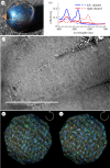
 has to be a multiple of the wavelength of light to satisfy the constructive interference condition. (Online version in colour.)


Similar articles
-
Structural colour and iridescence in plants: the poorly studied relations of pigment colour.Ann Bot. 2010 Apr;105(4):505-11. doi: 10.1093/aob/mcq007. Epub 2010 Feb 7. Ann Bot. 2010. PMID: 20142263 Free PMC article.
-
Function of blue iridescence in tropical understorey plants.J R Soc Interface. 2010 Dec 6;7(53):1699-707. doi: 10.1098/rsif.2010.0201. Epub 2010 Jun 2. J R Soc Interface. 2010. PMID: 20519208 Free PMC article.
-
Pointillist structural color in Pollia fruit.Proc Natl Acad Sci U S A. 2012 Sep 25;109(39):15712-5. doi: 10.1073/pnas.1210105109. Epub 2012 Sep 10. Proc Natl Acad Sci U S A. 2012. PMID: 23019355 Free PMC article.
-
Photonic structures in biology.Nature. 2003 Aug 14;424(6950):852-5. doi: 10.1038/nature01941. Nature. 2003. PMID: 12917700 Review.
-
Colourful cones: how did flower colour first evolve?J Exp Bot. 2020 Jan 23;71(3):759-767. doi: 10.1093/jxb/erz479. J Exp Bot. 2020. PMID: 31714579 Review.
Cited by
-
Structural colour in Chondrus crispus.Sci Rep. 2015 Jul 3;5:11645. doi: 10.1038/srep11645. Sci Rep. 2015. PMID: 26139470 Free PMC article.
-
Experimental degradation of helicoidal photonic nanostructures in scarab beetles (Coleoptera: Scarabaeidae): implications for the identification of circularly polarizing cuticle in the fossil record.J R Soc Interface. 2018 Nov 14;15(148):20180560. doi: 10.1098/rsif.2018.0560. J R Soc Interface. 2018. PMID: 30429263 Free PMC article.
-
Flower colours through the lens: quantitative measurement with visible and ultraviolet digital photography.PLoS One. 2014 May 14;9(5):e96646. doi: 10.1371/journal.pone.0096646. eCollection 2014. PLoS One. 2014. PMID: 24827828 Free PMC article.
-
Bioinspired phase-separated disordered nanostructures for thin photovoltaic absorbers.Sci Adv. 2017 Oct 20;3(10):e1700232. doi: 10.1126/sciadv.1700232. eCollection 2017 Oct. Sci Adv. 2017. PMID: 29057320 Free PMC article.
-
Development of structural colour in leaf beetles.Sci Rep. 2017 May 2;7(1):1373. doi: 10.1038/s41598-017-01496-8. Sci Rep. 2017. PMID: 28465577 Free PMC article.
References
-
- Vukusic P, Sambles JR, Lawrence CR, Wootton RJ. 1999. Quantified interference and diffraction in single Morpho butterfly scales. Proc. R. Soc. Lond. B 266, 1403–1411. (10.1098/rspb.1999.0794) - DOI
Publication types
MeSH terms
LinkOut - more resources
Full Text Sources
Other Literature Sources

