. 2013 Oct 27;96(8):689-96.
doi: 10.1097/TP.0b013e31829f1607.

Inhibitory effects of belatacept on allospecific regulatory T-cell generation in humans

Affiliations

Inhibitory effects of belatacept on allospecific regulatory T-cell generation in humans

Josh Levitsky et al. Transplantation. .

Abstract

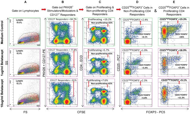
Background: It is unclear if new costimulatory blockade agents, such as the cytotoxic T lymphocyte-associated antigen 4-Ig molecule belatacept (BEL), promote or inhibit the potential for immunologic tolerance in transplantation. We therefore tested the in vitro effects of BEL on human regulatory T cells (Tregs) in mixed lymphocyte reactions (MLR) alone and in combination with maintenance agents used in transplant recipients.

Methods: BEL, mycophenolic acid (MPA), and sirolimus, either alone or in combination, were added to healthy volunteer Treg-MLR, testing (a) H-TdR incorporation for inhibition of lymphoproliferation and (b) flow cytometry to analyze for newly generated CD4+ CD25(high) FOXP3+ Tregs in carboxyfluorescein succinimidyl ester-labeled MLR responders. In addition, the modulatory effects of putative Tregs generated in the presence of these drugs were also tested using the lymphoproliferation and flow cytometric assays.

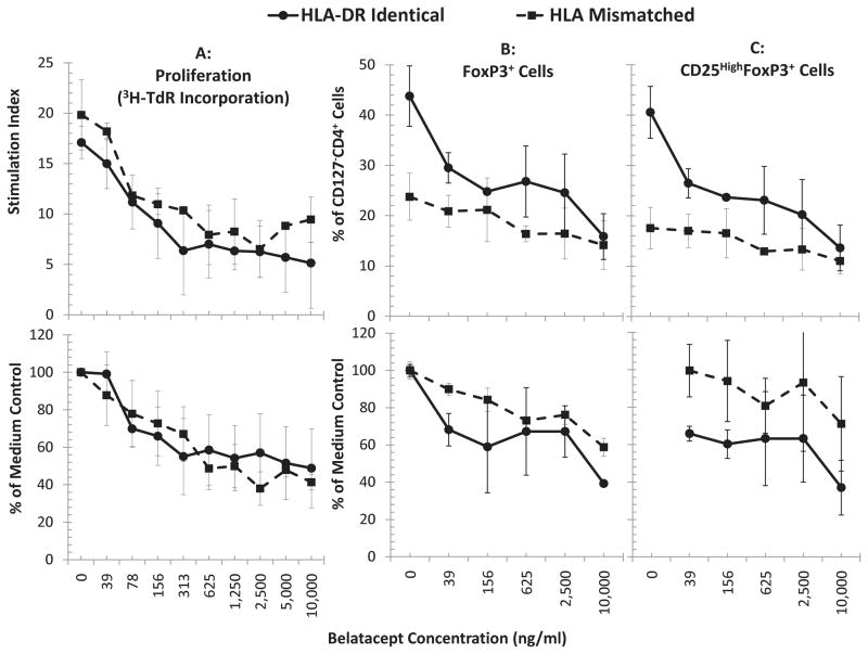
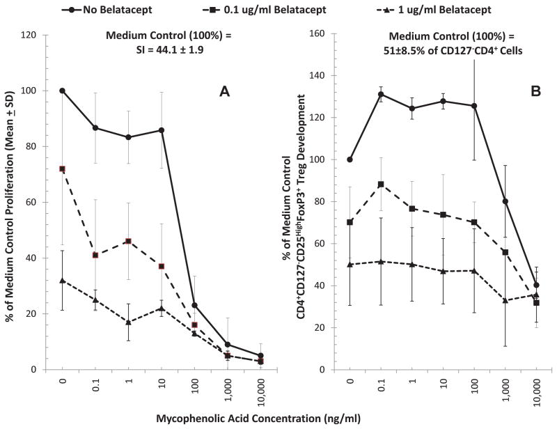
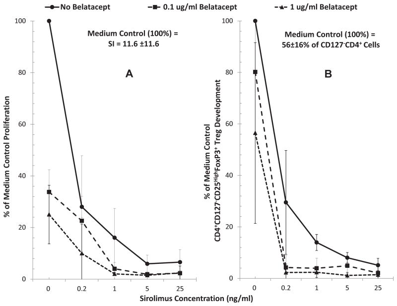
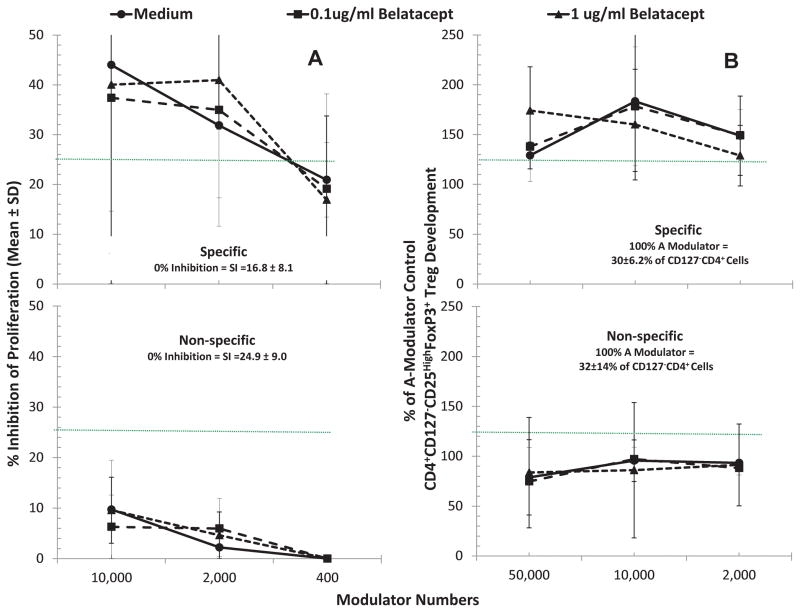
Results: In comparison with medium controls, BEL dose-dependently inhibited both lymphoproliferation and Treg generation in human leukocyte antigen DR matched and mismatched MLRs either alone or in combination with MPA or sirolimus. However, MPA alone inhibited lymphoproliferation but significantly enhanced Treg generation at subtherapeutic concentrations (P<0.01). In addition, purified CD4+ CD127- cells generated in MLR in the presence of MPA and added as third component modulators in fresh MLRs significantly enhanced newly developed Tregs in the proliferating responder cells compared with those generated with BEL or medium controls.

Conclusions: BEL alone and in combination with agents used in transplant recipients inhibits the in vitro generation of human Tregs. BEL might therefore be a less optimal agent for tolerance induction in human organ transplantation.


Figures

Figure 1
Figure 1. Scheme of flow analysis (representative 7-day experiment shown)
5x10⁵ CFSE labeled responding PBMC from healthy volunteer A were cultured with 5x10⁵ PKH26 labeled irradiated stimulator cells from laboratory volunteer B in the absence or presence of indicated concentrations of BEL. After 7 days, flow cytometric analyses were performed using monoclonal antibodies CD127-PE, CD4-ECD, CD25-PC7 and FOXP3-PC5. Viable lymphocytes were gated (column A) followed by CFSE bright and dim cells which were negative for either CD127-PE or PKH26 (column B), thus gating out CD127+ responders and any residual stimulators. This was followed by gating for CD4+ cells that were either non-proliferating (CFSE high) or proliferating (CFSE low) (column C). The cells in the non-proliferating (Column D) and proliferating (Column E) populations were analyzed by dot plots for CD25+ and FOXP3+ cells (among other subsets; not shown). Please note that when compared to the medium control (top row), fewer CD4+ cells proliferated in presence of BEL (column C). Additionally, there was a dose dependent reduction in the proportion of both total CD25+FOXP3+ Tregs and CD25highFOXP3+Tregs in the proliferating CD4+CD127 responder cells (column E). Since only the CFSE diluted proliferating fraction (as opposed to non-proliferating fraction; column D) demonstrated differences under various culture conditions, the results from only these are shown in subsequent experiments.
Figure 2
Figure 2. Effect of Belatacept on lymphoproliferation and Treg expansion in MLR (n=4):(B and C)
5x10⁵ CFSE labeled responding PBMC from healthy volunteer A were cultured with 5x10⁵ PKH26 labeled x-irradiated stimulator cells from HLA-DR identical or HLA-mismatched healthy volunteers (Bx or Ix respectively) in the presence of the indicated concentrations of BEL. On days 5, 7 and 9 flow cytometric analysis using the scheme shown in Figure 1 was performed and the percentage of CD4+CD127FOXP3+ cells (total Tregs; B) or CD4+CD127CD25HighFOXP3+ cells (natural Tregs; C) were quantitated. (A) In parallel, standard 3H thymidine incorporation assays were performed with 1x10⁵ PBMC each of the responders and stimulators in presence of indicated concentrations of BEL. The results from peak responses on day 7 are depicted. BEL inhibited the proliferation in 3H-thymidine incorporation assay (A); similarly BEL inhibited the generation of CD4+CD127FOXP3+ total-Tregs (B) and CD4+CD127CD25highFOXP3+ natural-Tregs (C) in a dose dependent manner (p<0.05, n=12). The data are shown as raw values (top row); and to minimize the variations between experiments, the data were also calculated as percentage of medium control (bottom row). In subsequent figures the data are shown as percentage of medium controls. Also, since similar profile was obtained for both total and natural Tregs, the data for only natural-Tregs are shown subsequently.
Figure 3
Figure 3. Effect of Belatacept and Mycophenolic Acid combinations on lymphoproliferation and Treg generation in MLR
Since BEL is used clinically in combination with MPA (the in vivo active form of MMF), MLRs were also performed in presence of combinations of these drugs. Both flow 3H-TdR uptake assays and flow cytometric analysis were performed, the latter as described in Figures 1 and 2, with serial dilutions of MPA as indicated on the horizontal axis and with 0.1μg/ml or 1μg/ml concentrations of BEL. (n=3). MPA increased the generation of Tregs at sub-therapeutic concentrations, while inhibiting lymphoproliferation (top blue line with no BEL) (p<0.05). BEL, however, not only inhibited lymphoproliferation and Treg generation (points at 0 MPA) as described in Figure 2, but also abrogated the enhancement of Treg generation observed at low concentrations of MPA.
Figure 4
Figure 4. Effect of Belatacept and Sirolimus combinations on lymphoproliferation and Treg generation in MLR
Since another drug that can potentially be used with BEL is SRL (with or without MMF), MLRs were also performed in presence of combinations of these drugs. Both flow 3H-TdR uptake assays and flow cytometric analysis were performed, the latter as described in Figures 1 and 2, with serial dilutions of SRL as indicated on the horizontal axis and with 0.1μg/ml or 1μg/ml concentrations of BEL (n=3). Both BEL SRL inhibited lymphoproliferation and Treg generation and the combination of the two had summative effect (p<0.05).
Figure 5
Figure 5. Assessment of CD4+ CD25highFOXP3+ in vitro inhibitory and recruitment functions by Tregs generated in MLR with or without Belatacept
Bulk cultures of responder PBMC from volunteer A were stimulated with 2 DR-matched x-irradiated cells from volunteer B either in the presence of no drugs (Medium), 0.1ug/ml or 1μg/ml BEL for 7 days. Then, after depleting CD127+ cells, the CD4+ cells were enriched as described in the methods. 5A: MLR Inhibition: Standard 3H thymidine incorporation assays were performed with 1x10⁵ each of the responders and stimulators in presence of indicated numbers of CD127CD4+ cells from the above bulk cultures with vs. without BEL, contrasted with control fresh A-PBMC modulators. Note that there appeared to be no greater effect of CD127CD4+ cells from “A+Bx+BEL” cultures in inhibiting 7-day 3H-TdR incorporation (not statistically significant) over that of CD127CD4+ cells immunoselected from “A+Bx+Medium” cultures (p>0.2). Nonetheless, there was a much less marked inhibition if the stimulator cells came from a non-specific donor (Ix) (p<0.05). 5B: Treg Recruitment: In parallel, 5x10⁵ CFSE labeled responding fresh PBMC from healthy volunteer A were cultured with 5x10⁵ PKH26-labeled stimulator cells from the original healthy volunteer B in the presence of the indicated numbers of PKH26-labeled CD127CD25+ modulators. On days 5, 7 and 9 flow cytometric analysis using the scheme shown in Figure 1 was performed (including the gating out of the PKH26 positive modulators and residual stimulators as well as the CD127+ responders). Data are calculated as percentage increase in CD4+CD25highFOXP3+ cell generation with the indicated modulators, over that observed with the control fresh A-PBMC modulators (100% shown by dotted line). CD127CD4+ cells from both “A+Bx+Medium” and “A+Bx+BEL” (allospecific) cultures similarly enhanced Treg generation (p>0.2). Also, as indicated in the lower panel, using an HLA-mismatched indifferent stimulator (Ix) virtually completely abrogated the in vitro recruitment effect seen in the allospecific cultures (p=0.02).

References

    1. Larsen CP, Pearson TC, Adams AB, et al. Rational development of LEA29Y (belatacept), a high-affinity variant of CTLA4-Ig with potent immunosuppressive properties. Am J Transplant. 2005;5 (3):443. - PubMed
    1. Durrbach A, Pestana JM, Pearson T, et al. A phase III study of belatacept versus cyclosporine in kidney transplants from extended criteria donors (BENEFIT-EXT study) Am J Transplant. 2010;10 (3):547. - PubMed
    1. Vincenti F, Charpentier B, Vanrenterghem Y, et al. A phase III study of belatacept-based immunosuppression regimens versus cyclosporine in renal transplant recipients (BENEFIT study) Am J Transplant. 2010;10 (3):535. - PubMed
    1. Levitsky J, Mathew JM, Flaa CW, Rosen A, Tambur AR, Miller J. The human “Treg MLR”: Immune monitoring for FOXP3+ T Regulatory Cell Generation. Transplantation. 2009 Dec;88(11):1303. - PMC - PubMed
    1. Szabo G, Gavala C, Mandrekar P. Tacrolimus and cyclosporine A inhibit allostimulatory capacity and cytokine production of human myeloid dendritic cells. Journal of Investigative Medicine. 2001;49 (5):442. - PubMed

Publication types

MeSH terms