Multicenter Study
. 2013 Aug 20;81(8):736-44.
doi: 10.1212/WNL.0b013e3182a1aafe. Epub 2013 Jul 24.

Impaired default network functional connectivity in autosomal dominant Alzheimer disease

Affiliations
Multicenter Study

Impaired default network functional connectivity in autosomal dominant Alzheimer disease

Jasmeer P Chhatwal et al. Neurology. .

Abstract

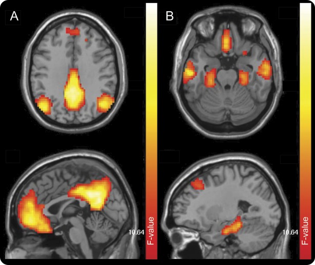
Objective: To investigate default mode network (DMN) functional connectivity MRI (fcMRI) in a large cross-sectional cohort of subjects from families harboring pathogenic presenilin-1 (PSEN1), presenilin-2 (PSEN2), and amyloid precursor protein (APP) mutations participating in the Dominantly Inherited Alzheimer Network.

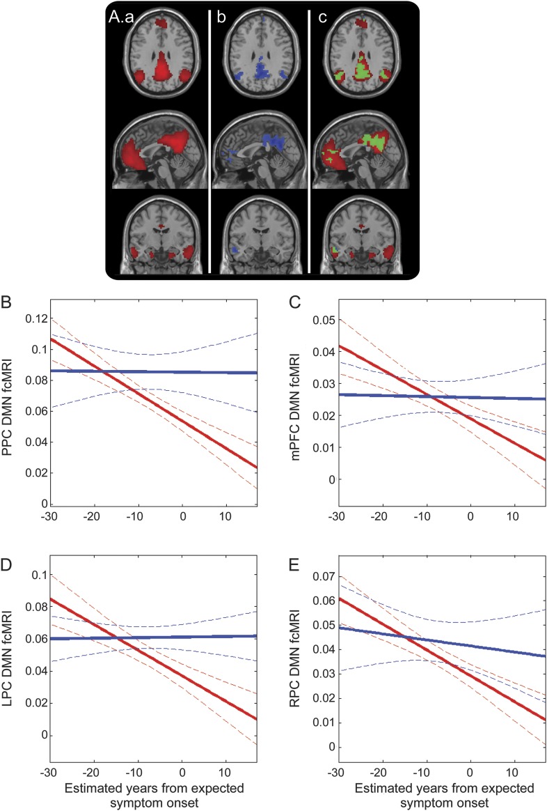
Methods: Eighty-three mutation carriers and 37 asymptomatic noncarriers from the same families underwent fMRI during resting state at 8 centers in the United States, United Kingdom, and Australia. Using group-independent component analysis, fcMRI was compared using mutation status and Clinical Dementia Rating to stratify groups, and related to each participant's estimated years from expected symptom onset (eYO).

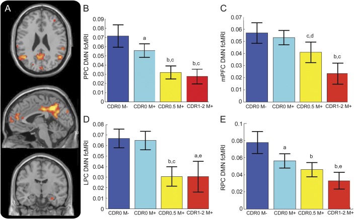
Results: We observed significantly decreased DMN fcMRI in mutation carriers with increasing Clinical Dementia Rating, most evident in the precuneus/posterior cingulate and parietal cortices (p < 0.001). Comparison of asymptomatic mutation carriers with noncarriers demonstrated decreased fcMRI in the precuneus/posterior cingulate (p = 0.014) and right parietal cortex (p = 0.0016). We observed a significant interaction between mutation carrier status and eYO, with decreases in DMN fcMRI observed as mutation carriers approached and surpassed their eYO.

Conclusion: Functional disruption of the DMN occurs early in the course of autosomal dominant Alzheimer disease, beginning before clinically evident symptoms, and worsening with increased impairment. These findings suggest that DMN fcMRI may prove useful as a biomarker across a wide spectrum of disease, and support the feasibility of DMN fcMRI as a secondary endpoint in upcoming multicenter clinical trials in Alzheimer disease.


Figures

Figure 1
Figure 1. DMN component derived from group ICA
The DMN was readily identified in the study population in one component of the group ICA. Dorsal (A) and ventral (B) portions of this component are shown above. DMN = default mode network; ICA = independent component analysis.
Figure 2
Figure 2. Whole-brain analysis of variance
(A) Whole-brain map depicting regions of significantly altered DMN fcMRI across groups (p < 0.001 threshold). Comparison across groups using a 10-mm sphere region of interest in the PPC (B), mPFC (C), LPC (D), and RPC (E). For post hoc analyses, error bars are SEM. a p < 0.05 compared with CDR 0M−; b p < 0.001 compared with CDR 0M−; c p < 0.001 compared with CDR 0M+; d p < 0.005 compared with CDR 0M−; e p < 0.05 compared with CDR 0M+. CDR = Clinical Dementia Rating; DMN = default mode network; fcMRI = functional connectivity MRI; LPC = left lateral parietal cortex; mPFC = medial prefrontal cortex; PPC = precuneus/posterior cingulate; RPC = right lateral parietal cortex.
Figure 3
Figure 3. Decreasing DMN connectivity as subjects approach and surpass their expected age of symptom onset
(A) Illustration of the DMN component (a; red); regions within the DMN that show negative correlation with eYO (p < 0.001; b; blue); overlap between the left and middle panels with common regions in green (c). Decreased connectivity within the DMN as measured in a 10-mm sphere based in the PPC (B), mPFC (C), LPC (D), and RPC (E). Solid lines denote linear correlation (with M− in blue and M+ in red) and curved dotted boundaries represent 95% confidence intervals. DMN = default mode network; eYO = estimated years from expected symptom onset; fcMRI = functional connectivity MRI; LPC = left lateral parietal cortex; mPFC = medial prefrontal cortex; PPC = precuneus/posterior cingulate; RPC = right lateral parietal cortex.

References

    1. Hardy J, Selkoe D. The amyloid hypothesis of Alzheimer's disease: progress and problems on the road to therapeutics. Science 2002;297:353–356 - PubMed
    1. Celone KA, Calhoun VD, Dickerson BC, et al. Alterations in memory networks in mild cognitive impairment and Alzheimer's disease: an independent component analysis. J Neurosci 2006;26:10222–10231 - PMC - PubMed
    1. Jones DT, Machulda MM, Vemuri P, et al. Age-related changes in the default mode network are more advanced in Alzheimer disease. Neurology 2011;77:1524–1531 - PMC - PubMed
    1. Greicius MD, Srivastava G, Reiss AL, Menon V. Default-mode network activity distinguishes Alzheimer's disease from healthy aging: evidence from functional MRI. Proc Natl Acad Sci USA 2004;101:4637–4642 - PMC - PubMed
    1. Fox MD, Snyder AZ, Vincent JL, Corbetta M, Van Essen DC, Raichle ME. The human brain is intrinsically organized into dynamic, anticorrelated functional networks. Proc Natl Acad Sci USA 2005;102:9673–9678 - PMC - PubMed

Publication types