

Botulinum toxin complex increases paracellular permeability in intestinal epithelial cells via activation of p38 mitogen-activated protein kinase
- PMID: 23884081
- PMCID: PMC3942962
- DOI: 10.1292/jvms.13-0164
Botulinum toxin complex increases paracellular permeability in intestinal epithelial cells via activation of p38 mitogen-activated protein kinase
Abstract
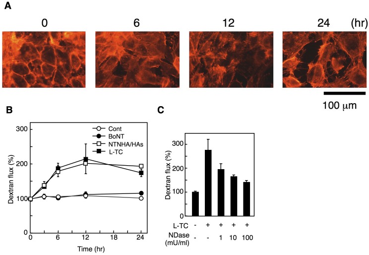
Clostridium botulinum produces a large toxin complex (L-TC) that increases paracellular permeability in intestinal epithelial cells by a mechanism that remains unclear. Here, we show that mitogen-activated protein kinases (MAPKs) are involved in this permeability increase. Paracellular permeability was measured by FITC-dextran flux through a monolayer of rat intestinal epithelial IEC-6 cells, and MAPK activation was estimated from western blots. L-TC of C. botulinum serotype D strain 4947 increased paracellular dextran flux and activated extracellular signal-regulated kinase (ERK), p38, but not c-Jun N-terminal kinase (JNK) in IEC-6 cells. The permeability increase induced by L-TC was abrogated by the p38 inhibitor SB203580. These results indicate that L-TC increases paracellular permeability by activating p38, but not JNK and ERK.
Figures
Similar articles
-
Differential role of Rho GTPases in intestinal epithelial barrier regulation in vitro.J Cell Physiol. 2011 May;226(5):1196-203. doi: 10.1002/jcp.22446. J Cell Physiol. 2011. PMID: 20945370
-
IKappaB-kinase/nuclear factor-kappaB signaling prevents thermal injury-induced gut damage by inhibiting c-Jun NH2-terminal kinase activation.Crit Care Med. 2007 May;35(5):1332-40. doi: 10.1097/01.CCM.0000261891.30360.F0. Crit Care Med. 2007. PMID: 17414734
-
[The role of mitogen-activated protein kinase cascades in inhibition of proliferation in human prostate carcinoma cells by raloxifene: an in vitro experiment].Zhonghua Yi Xue Za Zhi. 2008 Jan 22;88(4):271-5. Zhonghua Yi Xue Za Zhi. 2008. PMID: 18361842 Chinese.
-
Inhibition of p38 mitogen-activated protein kinase attenuates butyrate-induced intestinal barrier impairment in a Caco-2 cell monolayer model.J Pediatr Gastroenterol Nutr. 2014 Aug;59(2):264-9. doi: 10.1097/MPG.0000000000000369. J Pediatr Gastroenterol Nutr. 2014. PMID: 24625969
-
Modulation of renal epithelial barrier function by mitogen-activated protein kinases (MAPKs): mechanism of cyclosporine A-induced increase in transepithelial resistance.Kidney Int. 2003 Mar;63(3):908-16. doi: 10.1046/j.1523-1755.2003.00804.x. Kidney Int. 2003. PMID: 12631071
Cited by
-
GDNF is involved in the barrier-inducing effect of enteric glial cells on intestinal epithelial cells under acute ischemia reperfusion stimulation.Mol Neurobiol. 2014 Oct;50(2):274-89. doi: 10.1007/s12035-014-8730-9. Epub 2014 May 31. Mol Neurobiol. 2014. PMID: 24878766
References
-
- Bennett B. L., Sasaki D. T., Murray B. W., O’Leary E. C., Sakata S. T., Xu W., Leisten J. C., Motiwala A., Pierce S., Satoh Y., Bhagwat S. S., Manning A. M., Anderson D. W.2001. SP600125, an anthrapyrazolone inhibitor of Jun N-terminal kinase. Proc. Natl. Acad. Sci. U.S.A. 98: 13681–13686. doi: 10.1073/pnas.251194298 - DOI - PMC - PubMed
Publication types
MeSH terms
Substances
LinkOut - more resources
Full Text Sources
Other Literature Sources
Research Materials
Miscellaneous