

doi: 10.1038/jid.2013.312.
Epub 2013 Jul 24.
AKT1 gene mutation levels are correlated with the type of dermatologic lesions in patients with Proteus syndrome
Affiliations
- PMID: 23884311
- PMCID: PMC3868633
- DOI: 10.1038/jid.2013.312
Item in Clipboard
AKT1 gene mutation levels are correlated with the type of dermatologic lesions in patients with Proteus syndrome
J Invest Dermatol.
2014 Feb.
No abstract available
Figures
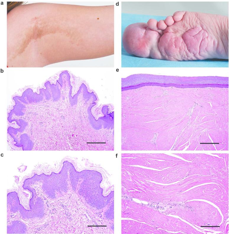
Representative examples of an EN and CCTN. Panel a shows the rough surface of the linear EN on the infraaxillary vault in patient 101. Hematoxylin and eosin stain (b, scale bar = 100 μm; c, scale bar = 50 μm) of a skin biopsy from the EN in panel a. The lesion is characterized by papillomatosis, mild hyperkeratosis, and acanthosis. Panel d shows the CCTN encompassing the hallux of patient 75. Hematoxylin and eosin stain of skin obtained after amputation of the hallux (e, scale bar = 100 μm; f, scale bar = 50 μm). Note the extensive dense collagen matrix, compression of the papillary dermis and the smooth surface of the lesion.
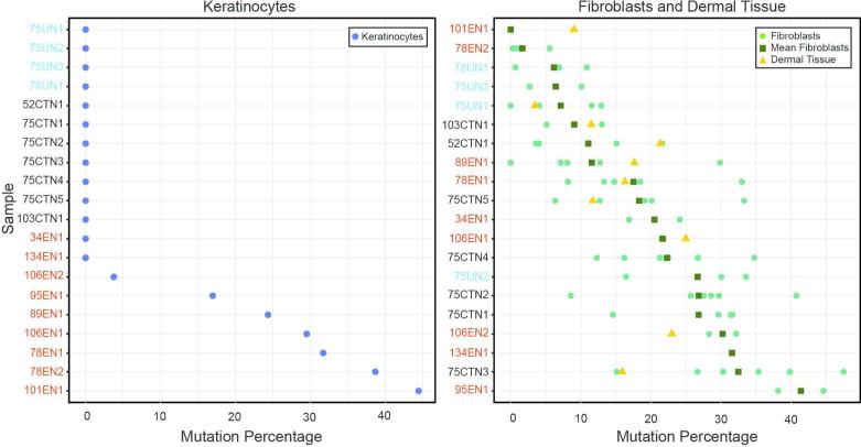
Percentage of AKT1 c.49G>A, p.Glu17Lys mutation in epidermal and dermal cells and tissue. One keratinocyte culture (left panel) was established from each epidermal sample. Mutation percentages are indicated by the blue circles. Multiple fibroblast cultures (right panel) were established from most pieces of dermal tissue. The mutation percentage for each fibroblast culture is indicated by a green circle. The mean mutation percentage for the fibroblast cultures from each sample is indicated by a green square and the mutation percentage in residual dermal tissue post culture is indicated by a yellow triangle. Samples are ordered from lowest to highest mutation value (keratinocytes) or mean mutation value (fibroblasts) and are color coded as follows: EN, red, CCTN, black, unaffected/unknown, light blue.
Similar articles
-
High-level somatic mosaicism of AKT1 c.49G>A mutation in skin scrapings from epidermal nevi enables non-invasive molecular diagnosis in patients with Proteus syndrome.Am J Med Genet A. 2013 Apr;161A(4):889-91. doi: 10.1002/ajmg.a.35764. Epub 2013 Feb 22. Am J Med Genet A. 2013. PMID: 23436452 No abstract available.
-
Epidermal Nevi and Related Syndromes -- Part 1: Keratinocytic Nevi.Actas Dermosifiliogr (Engl Ed). 2018 Oct;109(8):677-686. doi: 10.1016/j.ad.2018.05.005. Epub 2018 Jul 6. Actas Dermosifiliogr (Engl Ed). 2018. PMID: 29983155 Review. English, Spanish.
-
Late-onset Proteus syndrome with cerebriform connective tissue nevus and subsequent development of intraductal papilloma.Am J Med Genet A. 2022 Sep;188(9):2766-2771. doi: 10.1002/ajmg.a.62761. Epub 2022 Apr 20. Am J Med Genet A. 2022. PMID: 35441778 Free PMC article.
-
[A clinical study of Proteus syndrome caused by a mosaic somatic mutation in AKT1 gene].Zhonghua Nei Ke Za Zhi. 2019 Jul 1;58(7):508-513. doi: 10.3760/cma.j.issn.0578-1426.2019.07.005. Zhonghua Nei Ke Za Zhi. 2019. PMID: 31269567 Review. Chinese.
-
Progressive overgrowth of the cerebriform connective tissue nevus in patients with Proteus syndrome.J Am Acad Dermatol. 2010 Nov;63(5):799-804. doi: 10.1016/j.jaad.2009.12.012. Epub 2010 Aug 14. J Am Acad Dermatol. 2010. PMID: 20709429 Free PMC article.
Cited by
-
[Syndromes with vascular skin anomalies].Hautarzt. 2019 Jul;70(7):474-480. doi: 10.1007/s00105-019-4418-4. Hautarzt. 2019. PMID: 31111168 Review. German.
-
PIK3CA-related overgrowth spectrum (PROS): diagnostic and testing eligibility criteria, differential diagnosis, and evaluation.Am J Med Genet A. 2015 Feb;167A(2):287-95. doi: 10.1002/ajmg.a.36836. Epub 2014 Dec 31. Am J Med Genet A. 2015. PMID: 25557259 Free PMC article.
-
Repression of AKT signaling by ARQ 092 in cells and tissues from patients with Proteus syndrome.Sci Rep. 2015 Dec 11;5:17162. doi: 10.1038/srep17162. Sci Rep. 2015. PMID: 26657992 Free PMC article.
-
Pathogenetic insights from quantification of the cerebriform connective tissue nevus in Proteus syndrome.J Am Acad Dermatol. 2018 Apr;78(4):725-732. doi: 10.1016/j.jaad.2017.10.018. Epub 2017 Oct 16. J Am Acad Dermatol. 2018. PMID: 29042227 Free PMC article.
-
AKT kinases as therapeutic targets.J Exp Clin Cancer Res. 2024 Nov 29;43(1):313. doi: 10.1186/s13046-024-03207-4. J Exp Clin Cancer Res. 2024. PMID: 39614261 Free PMC article. Review.
References
-
- Aasen T, Belmonte JCI. Isolation and cultivation of human keratinocytes from skin or plucked hair for the generation of induced pluripotent stem cells. Nat Protoc. 2010;5:371–82. - PubMed
-
- Biesecker L. The challenges of Proteus syndrome: diagnosis and management. Eur J Hum Genet. 2006;14:1151–7. - PubMed
-
- Biesecker LG. The Multifaceted Challenges of Proteus Syndrome. JAMA. 2001;285:2240–3. - PubMed
-
- Groesser L, Herschberger E, Ruetten A, et al. Postzygotic HRAS and KRAS mutations cause nevus sebaceous and Schimmelpenning syndrome. Nat Genet. 2012;44:783–7. - PubMed
Publication types
MeSH terms
Substances
Supplementary concepts
Grants and funding
LinkOut - more resources
Full Text Sources
Other Literature Sources
Medical
Molecular Biology Databases
Miscellaneous
