. 2013 Sep 6;288(36):25804-25809.
doi: 10.1074/jbc.M113.474957. Epub 2013 Jul 24.

Ganglioside GM1-mediated transcytosis of cholera toxin bypasses the retrograde pathway and depends on the structure of the ceramide domain

Affiliations

Ganglioside GM1-mediated transcytosis of cholera toxin bypasses the retrograde pathway and depends on the structure of the ceramide domain

David E Saslowsky et al. J Biol Chem. .

Abstract

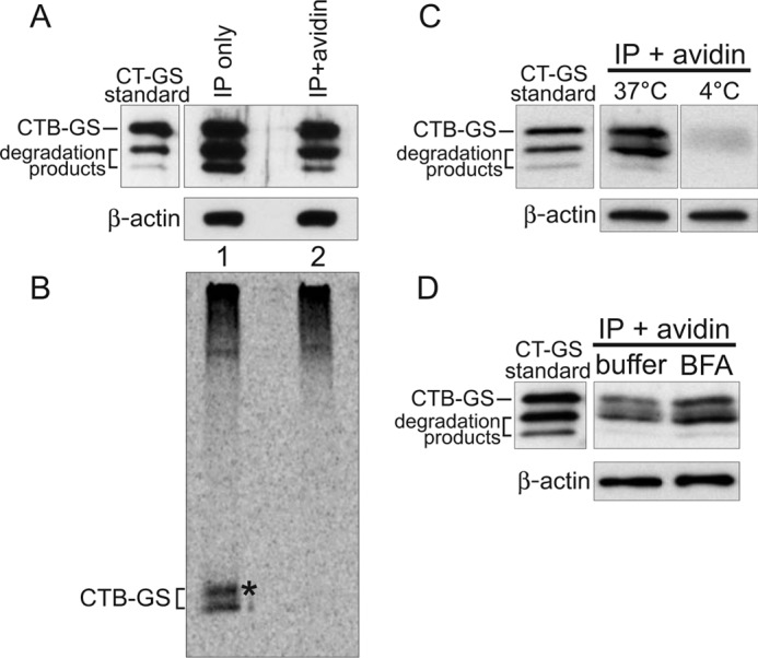
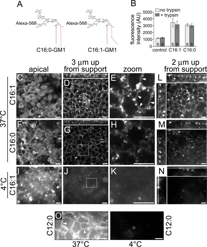
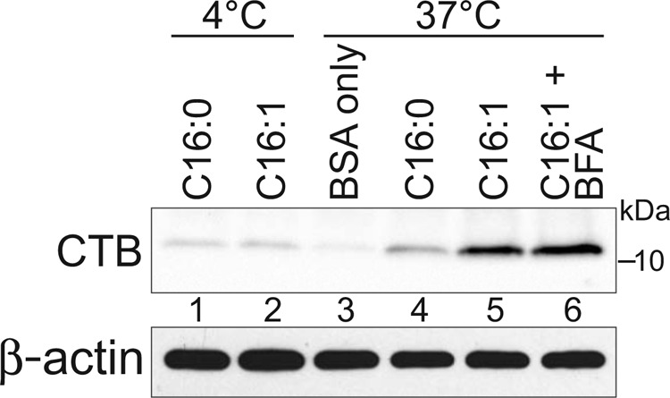
Cholera toxin causes diarrheal disease by binding ganglioside GM1 on the apical membrane of polarized intestinal epithelial cells and trafficking retrograde through sorting endosomes, the trans-Golgi network (TGN), and into the endoplasmic reticulum. A fraction of toxin also moves from endosomes across the cell to the basolateral plasma membrane by transcytosis, thus breeching the intestinal barrier. Here we find that sorting of cholera toxin into this transcytotic pathway bypasses retrograde transport to the TGN. We also find that GM1 sphingolipids can traffic from apical to basolateral membranes by transcytosis in the absence of toxin binding but only if the GM1 species contain cis-unsaturated or short acyl chains in the ceramide domain. We found previously that the same GM1 species are needed to efficiently traffic retrograde into the TGN and endoplasmic reticulum and into the recycling endosome, implicating a shared mechanism of action for sorting by lipid shape among these pathways.

Keywords: Cell Biology; Cholera Toxin; Endosomes; Ganglioside; Intracellular Trafficking; Lipid Transport; Membrane Lipids; Membrane Trafficking; Polarized Epithelia; Sphingolipid.


Figures

FIGURE 1.
FIGURE 1.
Sorting of CT into the transcytotic pathway bypasses retrograde transport to the TGN and ER. A–D, polarized T84 monolayers were incubated apically with 10 nm CT-GS for 2 h at 37 °C prior to biotinylation of the basolateral surfaces. Total toxin was immunoprecipitated from cell lysates and either analyzed directly (IP only, captures all CT-GS) or subjected to an additional avidin affinity purification step (IP + avidin, captures only basolateral CT-GS). To achieve similar CTB-GS levels between the total and basolateral pools, 7-fold more cells were used for the IP + avidin samples. 10% of each sample was analyzed by SDS-PAGE and immunoblot analysis using anti-CTB (A), with the remainder being separated by SDS-PAGE and analyzed by autoradiography (B). Pure CT-GS is indicated (standard), and crude lysates were separated by SDS-PAGE and probed with an antibody against β-actin as a loading control (A, C, and D, lower panels). The asterisk in B denotes a higher molecular weight glycosylated CTB-GS band. C, T84 monolayers were treated as in A, except cells were not pretreated with 35S-sulfate, and some cells were held at 4 °C during the 2-h apical toxin incubation. Basolateral CT-GS fractions (IP + avidin) were immunoblotted using anti-CTB. D, as in C, except cells were incubated with either buffer or 10 μm BFA for 20 min prior to apical exposure to 10 nm CT-GS for 2 h at 37 °C. Data are representative of three independent experiments for A–C and two independent experiments for D.
FIGURE 2.
FIGURE 2.
Polarized epithelial cells sort GM1 gangliosides into the basolaterally directed transcytotic pathway on the basis of the structure of the ceramide domain. A, schematic of GM1 structures. B, cell lysates of MDCK monolayers preincubated with the indicated Alexa Fluor-labeled GM1 species were measured by fluorimetry and reported as arbitrary units (AU) of fluorescence (± S.E.). The number of monolayers/condition is indicated at the column base. C–O, polarized epithelial monolayers were incubated apically with the indicated fluorophore-labeled GM1 species at 10 °C for 1 h, shifted to either 37 °C or 4 °C for the times indicated below, and imaged live by confocal microscopy. C–K, MDCK monolayers were incubated for 2 h at the indicated temperatures and imaged at the indicated Z plane (small panels above D and G show X-Z reconstructions). Zoomed images (E, H, and K) are from insets in D, G, and J. L–O, T84 monolayers were incubated apically with the indicated GM1 species for 3 h at the temperatures shown. Main panels in L-N are X-Y optical sections imaged 2 μm up from the basolateral support, and small panels at the top and left are X-Z and Y-Z reconstructions, respectively. O, T84 monolayers were incubated with Alexa Fluor-conjugated C12:0-GM1 at the indicated temperature and imaged 2 μm up from the basolateral support. Arrowheads indicate basolateral membrane labeling in E, L, and O. Scale bars = 10 μm. Data are representative of three independent experiments for B and O and at least six independent experiments for C–N.
FIGURE 3.
FIGURE 3.
Biochemical quantitation of C16:0/C16:1-GM1 transcytosis. Apical surfaces of MDCK monolayers were treated as indicated with C16:0-GM1 or C16:1-GM1 (no Alexa label, described under “Experimental Procedures”). CTB was immunoprecipitated from cell lysates, separated by SDS-PAGE, and immunoblotted using anti-CTB pAb (top panel). Pre-IP lysates were immunoblotted for β-actin as cell density control (bottom panel). Quantitation of CTB bands showed a 3.7- and 2.8-fold increase in lane 5 as compared with lane 4 in two independent experiments. The CTB band in lane 6 was 38 and 44% greater than lane 5 in two independent experiments. For these measurements, the relevant 4 °C CTB band (lanes 1 and 2) was considered as assay background and subtracted from the CTB bands in lanes 4–6 prior to comparison.

References

    1. Chinnapen D. J., Hsieh W. T., te Welscher Y. M., Saslowsky D. E., Kaoutzani L., Brandsma E., D'Auria L., Park H., Wagner J. S., Drake K. R., Kang M., Benjamin T., Ullman M. D., Costello C. E., Kenworthy A. K., Baumgart T., Massol R. H., Lencer W. I. (2012) Lipid sorting by ceramide structure from plasma membrane to ER for the cholera toxin receptor ganglioside GM1. Dev. Cell 23, 573–586 - PMC - PubMed
    1. Jobling M. G., Yang Z., Kam W. R., Lencer W. I., Holmes R. K. (2012) A single native ganglioside GM1-binding site is sufficient for cholera toxin to bind to cells and complete the intoxication pathway. mBio 3, e00401–12 - PMC - PubMed
    1. Wolf A. A., Jobling M. G., Saslowsky D. E., Kern E., Drake K. R., Kenworthy A. K., Holmes R. K., Lencer W. I. (2008) Attenuated endocytosis and toxicity of a mutant cholera toxin with decreased ability to cluster ganglioside GM1 molecules. Infect. Immun. 76, 1476–1484 - PMC - PubMed
    1. Lencer W. I., Moe S., Rufo P. A., Madara J. L. (1995) Transcytosis of cholera toxin subunits across model human intestinal epithelia. Proc. Natl. Acad. Sci. U.S.A. 92, 10094–10098 - PMC - PubMed
    1. Lencer W. I., Constable C., Moe S., Jobling M. G., Webb H. M., Ruston S., Madara J. L., Hirst T. R., Holmes R. K. (1995) Targeting of cholera toxin and E. coli heat labile toxin in polarized epithelia. Role of C-terminal KDEL. J. Cell Biol. 131, 951–962 - PMC - PubMed

Publication types