. 2013 Sep;140(17):3511-21.
doi: 10.1242/dev.098681. Epub 2013 Jul 24.

Notch/Rbpjκ signaling regulates progenitor maintenance and differentiation of hypothalamic arcuate neurons

Affiliations

Notch/Rbpjκ signaling regulates progenitor maintenance and differentiation of hypothalamic arcuate neurons

Paven K Aujla et al. Development. 2013 Sep.

Abstract

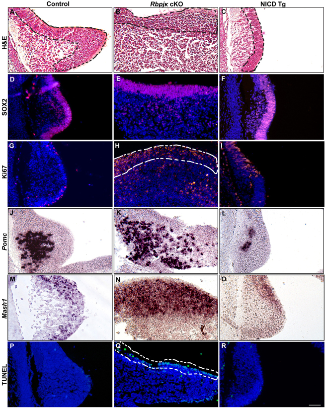
The hypothalamic arcuate nucleus (Arc), containing pro-opoiomelanocortin (POMC), neuropeptide Y (NPY) and growth hormone releasing hormone (GHRH) neurons, regulates feeding, energy balance and body size. Dysregulation of this homeostatic mediator underlies diseases ranging from growth failure to obesity. Despite considerable investigation regarding the function of Arc neurons, mechanisms governing their development remain unclear. Notch signaling factors such as Hes1 and Mash1 are present in hypothalamic progenitors that give rise to Arc neurons. However, how Notch signaling controls these progenitor populations is unknown. To elucidate the role of Notch signaling in Arc development, we analyzed conditional loss-of-function mice lacking a necessary Notch co-factor, Rbpjκ, in Nkx2.1-cre-expressing cells (Rbpjκ cKO), as well as mice with expression of the constitutively active Notch1 intracellular domain (NICD) in Nkx2.1-cre-expressing cells (NICD Tg). We found that loss of Rbpjκ results in absence of Hes1 but not of Hes5 within the primordial Arc at E13.5. Additionally, Mash1 expression is increased, coincident with increased proliferation and accumulation of Arc neurons at E13.5. At E18.5, Rbpjκ cKO mice have few progenitors and show increased numbers of differentiated Pomc, NPY and Ghrh neurons. By contrast, NICD Tg mice have increased hypothalamic progenitors, show an absence of differentiated Arc neurons and aberrant glial differentiation at E18.5. Subsequently, both Rbpjκ cKO and NICD Tg mice have changes in growth and body size during postnatal development. Taken together, our results demonstrate that Notch/Rbpjκ signaling regulates the generation and differentiation of Arc neurons, which contribute to homeostatic regulation of body size.

Keywords: Arcuate; Hypothalamus; Mouse; NPY; Notch; POMC.


Figures

Fig. 1.
Fig. 1.
Localization of Notch receptors and target genes in the developing ventral hypothalamus at E13.5. (A) Hematoxylin and Eosin staining on sagittal sections through the ventral midline at embryonic day 13.5 (E13.5) include the anterior ventral (AV) and posterior ventral (PV) hypothalamus. (B) In situ hybridization shows that Nkx2.1 mRNA is expressed in hypothalamic cells within the anterior and posterior ventral midline. (C-G) Pomc mRNA expression is detectable within the primordial arcuate nucleus (Arc; C). Additionally, Notch1 (D) and Notch2 (E) mRNAs are expressed in the AV hypothalamus, as well as Notch targets Hes1 (F) and Hes5 (G). Scale bar: 50 μm.
Fig. 2.
Fig. 2.
Loss of Rbpjκ as well as persistent expression of Notch1 within the developing Arc affects Hes1 expression and disrupts Arc morphology at E13.5. (A) Hematoxylin and Eosin staining on sagittal sections at the ventral midline include the primordial Arc (dashed box). (B,C) Rbpjκ cKO mice display an expansion (B) of the primordial Arc region (dashed boxes), whereas NICD Tg mice show a reduction (C) in Arc size. (D) In situ hybridization shows that Notch1 is expressed in the ventricular zone of the Arc (outlined). (E,F) Notch1 expression is maintained in this region in Rbpjκ cKO mice (E) and is abundantly expressed within the Arc progenitor region of NICD Tg mice (F). (G-I) Notch2 is also expressed in the within the Arc of control (G), Rbpjκ cKO (H) and NICD Tg (I) mice. (J-L) Hes1 is expressed within the control Arc (J), absent from Rbpjκ cKO mice (K) and expressed within an expanded region within the Arc of NICD Tg mice (L). (M-O) Surprisingly, Hes5 mRNA is expressed in the same region within the Arc of control animals (M), Rbpjκ cKO mice (N) and NICD Tg mice (O). Scale bar: 50 μm.
Fig. 3.
Fig. 3.
Notch signaling affects Arc progenitor population maintenance and Pomc differentiation at E13.5. (A-C) Hematoxylin and Eosin staining on Arc sagittal sections shows the Arc of control (A), Rbpjκ cKO (B) and NICD Tg (C) mice; dashed lines outline the more densely packed ventricular zone. (D-F) Immunohistochemistry probing expression of the progenitor marker Sox2 shows that cells within the ventricular zone are Sox2 positive within control (D), Rbpjκ cKO (E) and NICD Tg (F) mice. (G) Some cells within the Arc HVZ are Ki67 immunopositive in control mice. (H,I) Ki67-immunopositive cells are significantly increased in number in the Arc of Rbpjκ cKO mice (P<0.001; H) and within the NICD Tg Arc (P<0.001), but are restricted to a dorsal position (I). (J-L) Arc Pomc mRNA is detected in control mice (J). Rbpjκ cKO mice have a diffuse expression of Pomc-positive cells throughout the expanded Arc region (K), whereas NICD Tg mice have a reduction in Pomc-positive cells (L). (M-O) Similarly, Mash1 mRNA is detected within the Arc HVZ of control mice (M), diffusely expressed within the Arc of Rbpjκ cKO mice (N), and reduced in NICD Tg mice (O). (P,R) TUNEL staining reveals no cell death within the developing Arc in control (P) and NICD Tg mice (R). (Q) By contrast, TUNEL-positive cells within ventricular zone are found in Rbpjκ cKO mice (dashed lines). Scale bar: 50 μm.
Fig. 4.
Fig. 4.
Notch signaling is required for proper formation of the Arc hypothalamic ventricular zone at E18.5. (A) Hematoxylin and Eosin staining on coronal sections show the third ventricle (3V) and hypothalamic ventricular zone (HVZ; outlined). (B,C) Rbpjκ cKO mice show a loss of the Arc HVZ (B), whereas NICD Tg mice have an expanded Arc HVZ (C). (D-F) Sox2 immunostaining shows a loss of Sox2-immunopositive cells lining the 3V in Rbpjκ cKO mice (E) compared with controls (D), whereas NICD Tg animals have an expanded Arc HVZ comprising Sox2-immunopositive cells (F). (G-I) Nestin-immunopositive cells label neuronal progenitors along HVZ (G); this population is lost in Rbpjκ cKO mice (H) and expanded in NICD Tg mice (I). (J) In situ hybridization using the hypothalamic progenitor marker Rax labels cells within the Arc HVZ in controls animals. (K,L) Rbpjκ cKO mice show few Rax-positive cells (K), whereas NICD Tg animals have an expanded Rax-positive region (L). Scale bar: 50 μm.
Fig. 5.
Fig. 5.
Hypothalamic cells born at E12.5 remain in the hypothalamic ventricular zone when Notch is persistently expressed. (A,B) Coronal sections of the Arc at postnatal day 1 (P1) show that cells within the HVZ are not Ki67 immunopositive in control (A) or NICD Tg (B) mice, indicating that these cells are not proliferating at P1. BrdU immunohistochemistry was performed on mice at P1 that were injected with BrdU at E12.5. (C,D) BrdU-immunopositive cells are present within the HVZ and Arc (outlined) in control animals (C), but not in the Arc of NICD Tg mice (D). (E,F) Arc HVZ cells of NICD Tg mice are GFAP immunopositive (F), whereas control Arc HVZ cells do not express GFAP-immunopositive cells at this age (E). Scale bar: 50 μm.
Fig. 6.
Fig. 6.
Rbpjκ-dependent Notch signaling is required for proper differentiation of Arc neurons at E18.5. (A) In situ hybridization on coronal sections shows Pomc-positive cells in the Arc of controls. (B,C) Rbpjκ cKO mice have significantly more Pomc-positive neurons (B), whereas NICD Tg mice show no differentiation of Pomc-positive cells (C). (D-F) NPY-immunopositive cells are present in the Arc of control mice (D), are significantly increased in number (P<0.001; E) in Rbpjκ cKO animals and are not detected in NICD Tg animals (F). (G-I) In situ hybridization reveals Ghrh-positive cells in the Arc of control animals (G), an apparent increase in the number of these cells in Rbpjκ cKO mice (H) and no differentiation of these cells in NICD Tg mice (I). (J-L) Similarly, TH-immunopositive cells are present in the Arc of control mice (J) and appear increased in Rbpjκ cKO animals (K), whereas few cells are TH-immunopositive in NICD Tg mice (L). (M) Quantification of A-F (***P<0.001).
Fig. 7.
Fig. 7.
Body size is altered in mice with loss of Rbpjκ-dependent Notch signaling, as well as in mice with persistent Notch expression in the Arc. (A,B) Rbpjκ cKO mice appear normal in size and weight compared with controls at 2 postnatal weeks (2 wks; A), whereas NICD Tg mice are smaller in size (B). (C,D) At 8 weeks, Rbpjκ cKO (C) and NICD Tg (D) mice have increased body weight compared with littermate controls, yet NICD Tg mice appear shorter in length. Male and female mice were weighed once per week until 8 postnatal weeks; female mice are pictured. (E) Rbpjκ cKO mice begin increasing in body weight at week 5 and weigh significantly more than controls at week 7 (P<0.05) and week 8 (P<0.05). NICD Tg mice have significantly lower body weight at week 3 (**P<0.01), week 4 (*P<0.05) and week 5 (**P<0.01).
Fig. 8.
Fig. 8.
Notch/Rbpjκ signaling regulates progenitor maintenance and differentiation of hypothalamic Arc neurons through Hes1 activation and repression of Mash1.

References

    1. Acampora D., Postiglione M. P., Avantaggiato V., Di Bonito M., Vaccarino F. M., Michaud J., Simeone A. (1999). Progressive impairment of developing neuroendocrine cell lineages in the hypothalamus of mice lacking the Orthopedia gene. Genes Dev. 13, 2787–2800 - PMC - PubMed
    1. Akazawa C., Sasai Y., Nakanishi S., Kageyama R. (1992). Molecular characterization of a rat negative regulator with a basic helix-loop-helix structure predominantly expressed in the developing nervous system. J. Biol. Chem. 267, 21879–21885 - PubMed
    1. Akimoto M., Nishimaki T., Arai Y., Uchinuma E., Yamauchi H., Kameda Y. (2010). Hes1 regulates formations of the hypophyseal pars tuberalis and the hypothalamus. Cell Tissue Res. 340, 509–521 - PubMed
    1. Altman J., Bayer S. A. (1978). Development of the diencephalon in the rat. I. Autoradiographic study of the time of origin and settling patterns of neurons of the hypothalamus. J. Comp. Neurol. 182, 945–971 - PubMed
    1. Alvarez-Bolado G., Paul F. A., Blaess S. (2012). Sonic hedgehog lineage in the mouse hypothalamus: from progenitor domains to hypothalamic regions. Neural Dev. 7, 4 - PMC - PubMed

Publication types

MeSH terms

Substances