Review
. 2013:790:95-127.
doi: 10.1007/978-1-4614-7651-1_6.

Paramyxovirus entry

Affiliations
Review

Paramyxovirus entry

Katharine N Bossart et al. Adv Exp Med Biol. 2013.

Abstract

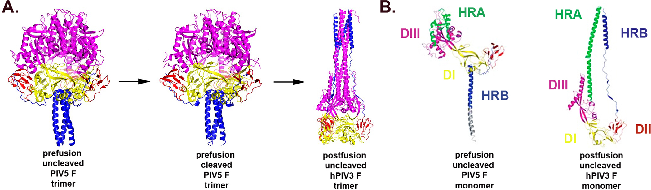
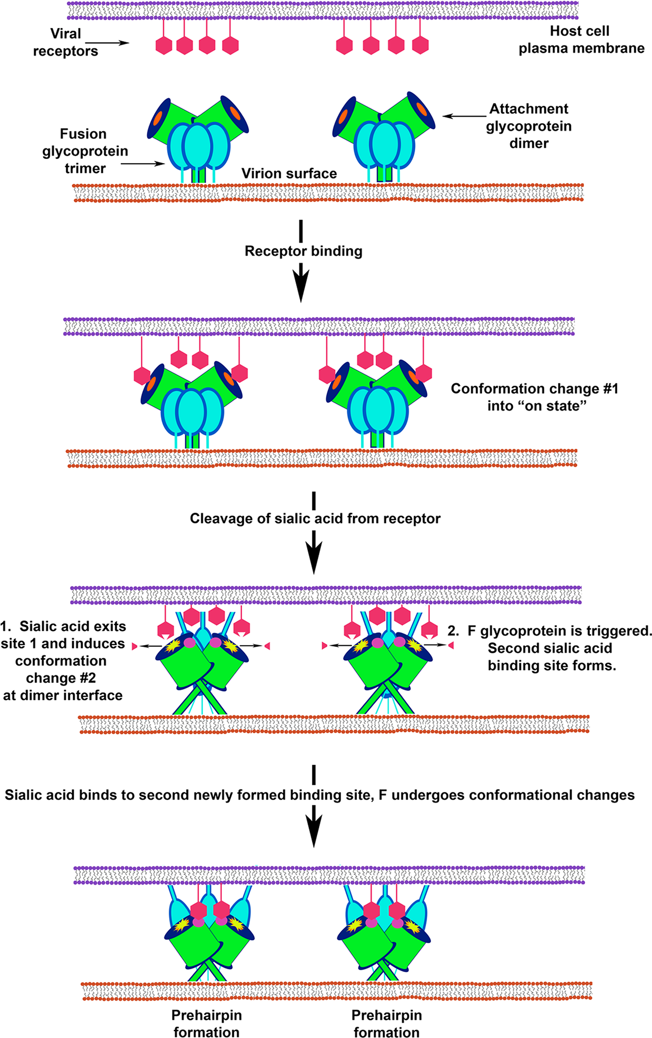
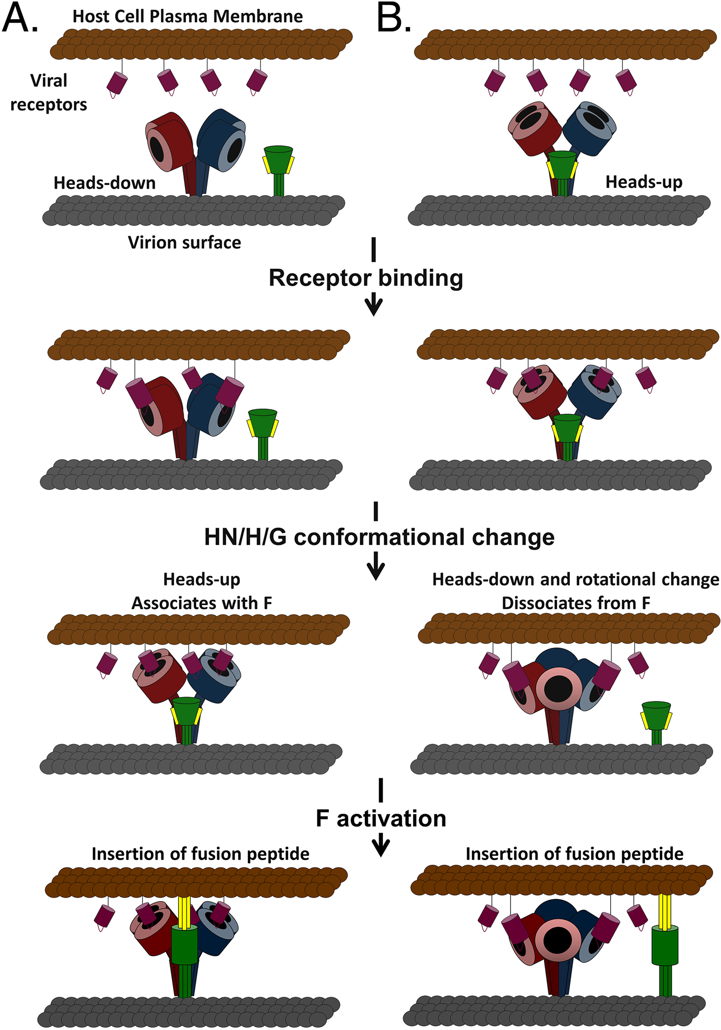
The family Paramyxoviridae consists of a group of large, enveloped, negative-sense, single-stranded RNA viruses and contains many important human and animal pathogens. Molecular and biochemical characterization over the past decade has revealed an extraordinary breadth of biological diversity among this family of viruses. Like all enveloped viruses, paramyxoviruses must fuse their membrane with that of a receptive host cell as a prerequisite for viral entry and infection. Unlike most other enveloped viruses, the vast majority of paramyxoviruses contain two distinct membrane-anchored glycoproteins to mediate the attachment, membrane fusion and particle entry stages of host cell infection. The attachment glycoprotein is required for virion attachment and the fusion glycoprotein is directly involved in facilitating the merger of the viral and host cell membranes. Here we detail important functional, biochemical and structural features of the attachment and fusion glycoproteins from a variety of family members. Specifically, the three different classes of attachment glycoproteins are discussed, including receptor binding preference, their overall structure and fusion promotion activities. Recently solved atomic structures of certain attachment glycoproteins are summarized, and how they relate to both receptor binding and fusion mechanisms are described. For the fusion glycoprotein, specific structural domains and their proposed role in mediating membrane merger are illustrated, highlighting the important features of protease cleavage and associated tropism and virulence. The crystal structure solutions of both an uncleaved and a cleavage-activated metastable F are also described with emphasis on how small conformational changes can provide the necessary energy to mediate membrane fusion. Finally, the different proposed fusion models are reviewed, featuring recent experimental findings that speculate how the attachment and fusion glycoproteins work in concert to mediate virus entry.


Figures

Figure 1.
Figure 1.. Negatively stained paramyxovirus virions.
A. Newcastle disease virus (avulavirus). B. Human parainfluenza virus type 3 (respirovirus). C. Hendra virus (henipavirus). D. Canine distemper virus (morbillivirus). E. Menangle virus (proposed rubulavirus). F. Respiratory syncytial virus (pneumovirus). G. J-virus (proposed jeilongvirus). H. Mossman virus (unclassified). All micrographs were adjusted to the same magnification with exception of panels F and G. For all panels: bar, 200nm. Images courtesy of the AAHL Biosecurity Microscopy Facility, Australian Animal Health Laboratory (AAHL) Livestock Industries CSIRO, Australia.
Figure 2.
Figure 2.. The attachment and fusion envelope glycoproteins.
A. Important functional domains. For the attachment glycoprotein the transmembrane domain, cytoplasmic tail, proposed stalk domain and globular head are indicated. For the fusion glycoprotein the F1 and F2 subunits are depicted. F1 contains the signal sequence, transmembrane domain, cytoplasmic tail, fusion peptide, heptad repeat A (HRA) and heptad repeat B (HRB). The important domains that constitute the globular head of the F trimer, DI, DII and DIII, are represented by different shading. B. Orientation of the attachment and fusion glycoprotein in the virion membrane. F is a typical type I membrane glycoprotein with one membrane spanning domain and an extracellular N-terminus. The disulfide bond that links the F1 and F2 subunits is also shown. The attachment glycoproteins are type II membrane proteins where the molecule’s amino (N)-terminus is oriented towards the cytoplasm and the protein’s carboxy (C)-terminus is extracellular.
Figure 3.
Figure 3.
(A) Structure of the NDV HN, Australia–Victoria (AV) strain ectodomain. Two dimers of the NDV HN NA domains flank the 4HB (residues 83–114) in the stalk. The four NA active sites are shown as blue spheres. The secondary sialic acid binding sites located at the NA domain dimer interface are shown as orange spheres. Mutations of NDV HN stalk residues R83, A89, L90, L94 and L97 are known to impair F activation specifically and are implicated in forming direct contacts with the F glycoprotein. These mutations reside along the stalk region marked by the arrow in the HN tetramer structure. (B) Model of HeV G. Left: The G ectodomain is shown in the dimer conformation with the two globular head domains derived from the crystal structure, colored in green and blue, with predicted N-linked glycosylation sites shown as gray spheres. The G head domain folds as a six-bladed β-propeller with disulfide bonds illustrated as yellow sticks. The residues of the ephrin-B2 G-H loop are also shown in yellow occupying the RBS. Stalk residues 77–136 are modeled for each monomer, and the position of the HeV G head dimer and stalks are oriented based on the alignment with the NDV structure. Ile residues in the HeV G stalk domain that modulate G fusion promotion activity are indicated. Right: Model of the HeV G dimer with globular heads and stalk domains as on right and rotated with residues G449 and D468 highlighted in red showing their proximity to the stalk domain. Mutation of these residues decreases HeV fusion, suggesting they may be involved in interactions between the globular heads and stalk domains that are essential for the fusion process. Figures have been modified from original work with permission from R.A. Lamb and Proceedings of the National Academy of Sciences and the creative commons public license.
Figure 4.
Figure 4.. Model showing how paramyxovirus F heptad repeats mediate membrane merger.
For simplicity, the attachment glycoprotein and its proposed role in triggering or promoting F activity is not shown. Upon activation, significant conformational changes in F lead to HRA forming a three-stranded α-helical coiled coil and the translocation of the fusion peptide and its intercalation into the target cell membrane. This fusion intermediate conformation of F is referred to as the prehairpin structure. Further conformational changes lead to the packing of α-helical HRB domains into the grooves of the HRA coiled coil in an antiparallel orientation giving rise to six-helix bundle formation. As the six-helix bundle forms, the two membranes are drawn closer together, and the energy released as F transitions to its most stable conformation is thought to drive membrane merger.
Figure 5.
Figure 5.. Structural changes between the pre- and postfusion F glycoprotein conformations derived from PIV5 and hPIV3.
A. Ribbon diagrams of uncleaved and cleavage-activated prefusion forms and postfusion F trimers are shown side by side. The three domains, DI, DII and DIII, are depicted by different shading (DI yellow, DII red and DIII pink). B. Ribbon diagrams of pre- and postfusion F monomers similarly oriented by DI as shown in Panel A. Monomers are shown side by side to demonstrate more clearly the different domains, DI, DII and DIII, which are depicted by different shading as in A. Adapted with permission from R.A. Lamb and Macmillan Publishers Ltd: Nature, and Proceedings of the National Academy of Sciences.
Figure 6.
Figure 6.. Multiple conformational changes in NDV HN trigger F from metastable to fusogenic.
Fusion model as described in Zaitsev et al depicting the receptor-triggered mechanism of NDV fusion. Receptor binding induces conformational changes that convert HN to its “on state”. Subsequent cleavage and release of sialic acid leads to conformational changes at the HN dimer interface that not only are critical for triggering F but also generate a second sialic acid binding site. Binding cellular receptors via the newly formed second sialic acid site is hypothesized to keep the virion in close proximity to the target cell membrane. For simplicity F is shown as a trimer with limited conformational changes and membrane merger has been excluded.
Figure 7.
Figure 7.. Models of paramyxovirus membrane fusion involving heads-up and heads-down conformations.
Initial expression of the tetrameric attachment (HN/H/G) glycoprotein (dimers colored red and blue) and the fusion (F) glycoprotein (green) is depicted in the (A) heads-down, non-F-associated or (B) heads-up, F-associated conformations. In both models, HN/H/G binds receptor (maroon) and undergoes receptor-induced conformational changes, switching from heads-up to heads-down or vice versa. The change in the position of the globular heads allows for (A) association (provocateur model) or (B) dissociation (clamp model) with F, leading to the fusion activation of F and the beginning of membrane fusion by the insertion of the fusion peptide (yellow) into the target cell membrane.

References

    1. Lamb RA, and Parks GD. Paramyxoviridae: The viruses and their replication. In Knipe DM and Howley PM ed., Fields Virology, 5 ed, Philadelphia. Lippincott Williams & Wilkins, 2007:1449–1496.
    1. Alexander DJ. Newcastle disease and other avian paramyxoviruses. Rev Sci Tech 2000; 19(2):443–462. - PubMed
    1. Tambi EN, Maina OW, Mukhebi AW, et al. Economic impact assessment of rinderpest control in Africa. Rev Sci Tech 1999; 18(2):458–477. - PubMed
    1. Eaton BT, Broder CC, Middleton D, et al. Hendra and Nipah viruses: different and dangerous. Nat Rev Microbiol 2006; 4(1):23–35. - PMC - PubMed
    1. Eaton BT, Mackenzie JS, and Wang L-F. Henipaviruses. In Knipe DM and Howley PM ed., Fields Virology, 5 ed, Philadelphia. Lippincott Williams & Wilkins, 2007:1587–1600.

Publication types

MeSH terms