. 2013 Aug 1;27(15):1680-92.
doi: 10.1101/gad.220095.113. Epub 2013 Jul 24.

Chromatin-to-nucleoprotamine transition is controlled by the histone H2B variant TH2B

Affiliations

Chromatin-to-nucleoprotamine transition is controlled by the histone H2B variant TH2B

Emilie Montellier et al. Genes Dev. .

Abstract

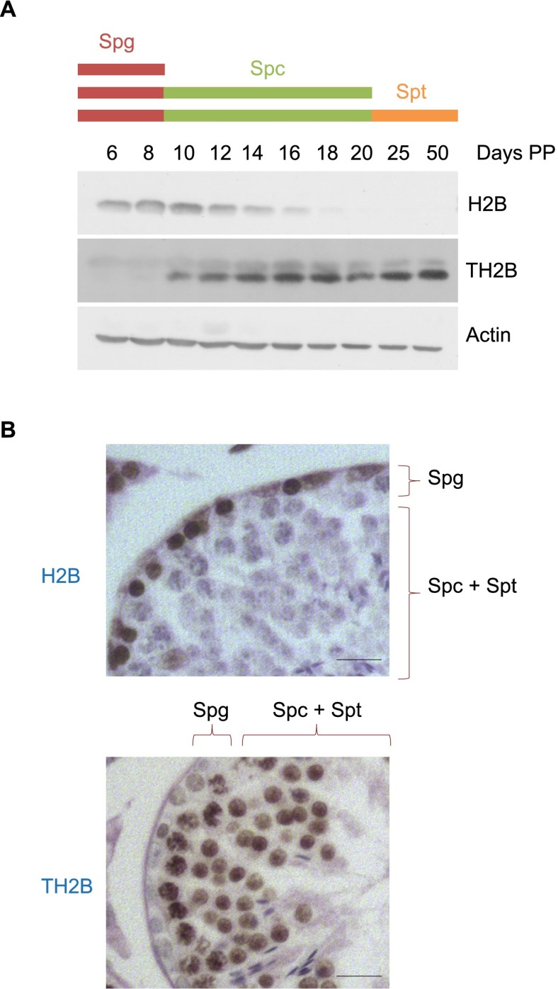
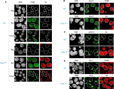
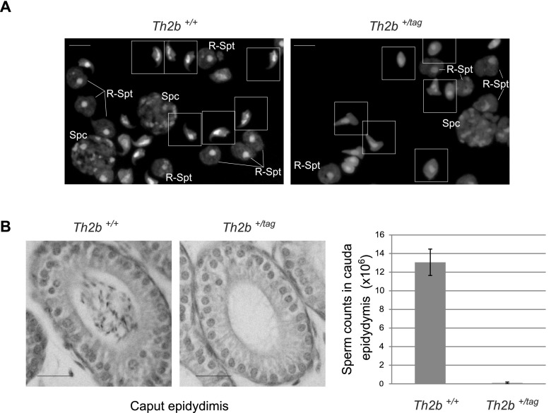
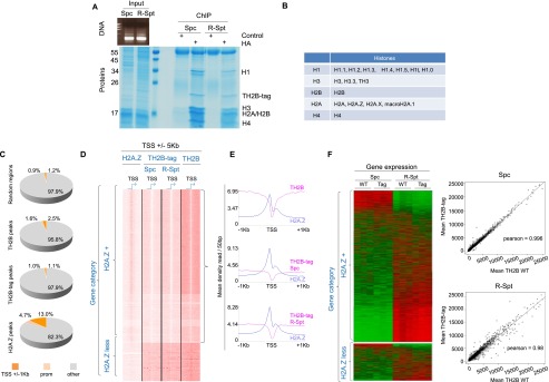
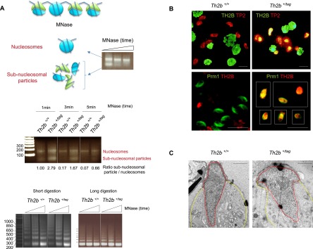
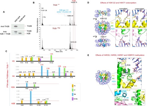
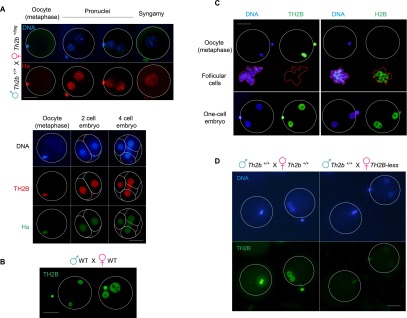
The conversion of male germ cell chromatin to a nucleoprotamine structure is fundamental to the life cycle, yet the underlying molecular details remain obscure. Here we show that an essential step is the genome-wide incorporation of TH2B, a histone H2B variant of hitherto unknown function. Using mouse models in which TH2B is depleted or C-terminally modified, we show that TH2B directs the final transformation of dissociating nucleosomes into protamine-packed structures. Depletion of TH2B induces compensatory mechanisms that permit histone removal by up-regulating H2B and programming nucleosome instability through targeted histone modifications, including lysine crotonylation and arginine methylation. Furthermore, after fertilization, TH2B reassembles onto the male genome during protamine-to-histone exchange. Thus, TH2B is a unique histone variant that plays a key role in the histone-to-protamine packing of the male genome and guides genome-wide chromatin transitions that both precede and follow transmission of the male genome to the egg.

Keywords: BRDT; H2AZ; histone eviction; male contraception; male infertility; reprogramming; sex chromosome inactivation.


Figures

Figure 1.
Figure 1.
A major H2B-to-TH2B transition occurs in early spermatocytes. (A) TH2B and H2B accumulation was analyzed in testes extracts at the indicated times (in days post-partum [Days PP]) by Western blots. (B) The expression of TH2B and H2B was analyzed by immunohistochemistry. Testis sections at stage II/III are represented. Spermatogonia (Spg) and spermatocytes and spermatids (Spc + Spt) are indicated. Bars, 10 μm.
Figure 2.
Figure 2.
TH2B and TH2B-tag show similar intracellular distribution in wild-type and TH2B-tag-expressing spermatogenic cells. (A) Codetection of TH2B and TH2B-tag was performed by immunofluorescence on seminiferous tubule preparations from testes from the indicated genotypes using anti-TH2B or anti-Ha antibodies. The stages considered are indicated at the left of each panel: spermatogonia (Spg), pachytene spermatocytes (Spc) and round spermatids (R-Spt). (B) Sycp3 was codetected with TH2B (top panel) or TH2B-tag (anti-Ha, bottom panel) in spermatocytes of the indicated genotypes. (C) Codetection of TH2B-tag (anti-Ha panels) and γH2A.X in spermatocytes with the indicated genotypes is shown. (D) H3.3 was codetected with TH2B or TH2B-tag in spermatocytes of the indicated genotypes. Please note that the immunodetection with anti-Ha antibody is more sensitive than the detection based on the anti-TH2B antibody.
Figure 3.
Figure 3.
TH2B-tag induces late arrest of spermatogenesis. (A) Different cell types stained with DAPI from the indicated genotypes are shown. Bars, 5 μm. (B) Cauda epididymis sections stained with hematoxylin are shown in the left panel, and spermatozoa counts from isolated cauda epididymis from mice with the indicated genotypes are presented as histograms in the right panel. Bars, 20 μm. Bars represent standard deviations of sperm counts from cauda epididymis of five mice of each genotype.
Figure 4.
Figure 4.
TH2B-tag is assembled into nucleosomes in spermatocytes and round spermatids and does not affect any of the fine-tuned chromatin activities. (A) Chromatin from spermatocytes (Spc) and round spermatids (R-Spt) was extensively digested with MNase (DNA gel) and subjected to immunoprecipitation using an anti-Ha antibody. The materials obtained from ChIP with anti-Ha or an irrelevant antibody were visualized after SDS-PAGE followed by Coomassie staining. The position of histones identified by MS is indicated. (B) The different variants found associated with TH2B nucleosomes are indicated (see also Supplemental Table S1). All of the unique peptides identified corresponding to each of the linker histone types are shown in Supplemental Table S2. (C) Mononucleosomes from cells isolated from Th2b+/tag (spermatocytes and round spermatids) or Th2b+/+ (total spermatogenic cell suspension) mouse testes were subjected to ChIP with anti-Ha and anti-TH2B antibodies, respectively, followed by DNA sequencing (ChIP-seq). The respective proportions of TH2B-tag and TH2B peaks (this experiment) distributed on gene regulatory regions (color-coded), including TSSs (regions covering TSSs ±1 kb) and promoter regions (from TSSs to 5 kb upstream), were compared with that of H2A.Z peaks (from spermatogenic cells, GSE29913) as well as with “random” genomic localizations (using a set of 75,000 200-bp segment regions randomly selected from the input raw read files). (D) SeqMiner software (Ye et al. 2011) was used to compare TH2B-tag's, TH2B's, and H2A.Z's respective distribution around the TSSs (±1 kb) of the same genes in the indicated spermatogenic cell populations ([Spc] spermatocytes; [R-Spt] spermatids) for the TH2B-tag and total spermatogenic cell population for TH2B. The genes were classified as either H2A.Z-positive (H2A.Z+) or H2A.Z-negative (H2A.Z-less). (E) Metagene analysis of TH2B-tag (in spermatocytes [Spc] and spermatids [R-Spt]) and TH2B (in total wild-type spermatogenic cells) together with H2A.Z distribution centered around the TSSs of the H2A.Z+ gene group. (F, left panel) Whole-genome gene expression pattern of Th2b+/+ (WT) and Th2b+/tag (Tag) spermatocytes (Spc) and round spermatids (R-Spt) are shown as heat maps for the H2A.Z+ and H2A.Z-less gene classes. (Right panel) Pearson correlation plots between individual gene expression in spermatocytes (Spc) and round spermatids (R-Spc), wild-type or expressing TH2B-tag, are shown.
Figure 5.
Figure 5.
TH2B-tag affects subnucleosomal transitional states in elongating spermatids during histone replacement. (A, top panel) Nuclei isolated from elongated/condensing wild-type spermatids were digested with MNase for increasing lengths of time to release both nucleosomal MNase-resistant regions and MNase-sensitive subnucleosomal particles (histone and nonhistone proteins are represented in blue and green, respectively). (Middle panel) Nuclei from wild-type elongated/condensing spermatids or the corresponding cells isolated from TH2B-tag-expressing spermatogenic cells were extensively digested by MNase as above. The agarose gel shows nucleosomal and subnucleosomal DNA fragments released from the two cell types by MNase digestion during the indicated times. The normalized ratios of intensity of subnucleosomal particles to nucleosomes obtained with ImageJ software are indicated below each lane. This ratio was set to 1 for Th2b+/+ after 1 min of MNase digestion and was used to normalize the other values. The bottom panels show chromatin digestion by MNase of suspensions of total spermatogenic cells from wild-type and TH2B-tag mouse testes. (B) Codetection of TH2B (green) and TP2 (red) (top panel) and of Th2B (red) and protamine 1 (Prm1, green) (bottom panel) were performed on spermatogenic cell preparations from wild-type or Th2b+/tag testes by immunofluorescence using the corresponding antibodies. Bars, 10 μm. (C) Electron micrographs show representative spermatids of both genotypes. Nucleus and cytoplasm are delimited by red and yellow lines, respectively. Bars, 1 μm.
Figure 6.
Figure 6.
A highly specific compensation mechanism is activated in the absence of TH2B. (A) Histone extracts from wild-type and TH2B-less spermatogenic cells were subjected to electrophoresis on TAU gels to allow separation of TH2B and H2B and were subsequently used for the immunodetection of the two histones by Western blotting with a specific anti-TH2B antibody (top panel) and an antibody recognizing both TH2B and H2B (bottom panel). (B) Profiling of histones from wild-type and TH2B-less testes was performed by UHPLC-MS. The figure shows the deconvoluted electrospray mass spectra of H4, TH2B, and H2B (see Supplemental Fig. S5A,B for additional information). (C) Histones from wild-type and TH2B-less testes were subjected to in vitro isotopic labeling followed by HPLC/MS/MS analysis of histone peptides. The relative abundance of the identified histone PTMs is expressed as TH2B-less/wild-type ratio (for details see Supplemental Table S4; Supplemental Fig. S5D). Lysine acetylation (Ac), crotonylation (Cr), and dimethylation (Di-Me) and arginine monomethylation (Me) are color-coded as indicated. (D) Lysine residues exhibiting enhanced crotonylation. (Left) Nucleosome core particle (Protein Data Bank [PDB] code 3AZH). Histones H2A, H2B, H3, and H4 are in cyan, pink, yellow, and green, respectively; DNA is in blue. Modified lysines are shown as red spheres. The dyad axis is vertical in the middle view. (Insets) Close-up of H3K122 (top) and H4K77 (bottom). Hypothetical crotonyl groups are also shown (blue ovals). (E) Arginine residues displaying enhanced methylation. (Left) Nucleosome showing modified arginines as red spheres. (Top inset) Close-up of H4R35 and H4R55. H4R35 forms a direct hydrogen (H) bond to the DNA backbone by adopting an orientation that is itself stabilized by an H bond with H4 residue Tyr51 (not shown). Disruption of either H bond by methylation would weaken DNA binding. H4R55 is in an intricate H bond network with H4 residues Gln27 and Glu52 and the backbone carbonyl of Ile29. This network orients a stretch of N-terminal H4 residues (residues 25–29) such that residue Asn25 is ideally placed to H-bond with H3 residue Glu73. R55 methylation would likely destabilize this network and perturb the precise orientation of the H4 N-terminal stretch, thereby weakening the H3–H4 interface. (Bottom inset) Close-up of H4R67 and H2BR72. Residue H4R67 is stabilized by an H bond with Asn64 in a conformation allowing it to form a cation–π interaction with H3 residue Phe78. Methylation would destabilize the latter interaction by disrupting the H bond and/or delocalizing the positive charge on the R67 guanidino group due to the electron-releasing inductive effect of the added methyl group. H2BR72 is in an H bond network with H2B residue Asp68, H4 residue Tyr98, and the backbone carbonyl of H2A residue Leu96. Methylation would disrupt this network and thus destabilize the H2B–H2A and/or H2B–H4 interfaces.
Figure 7.
Figure 7.
TH2B is assembled on the male genome at fertilization. (A, top panel) Th2b+/tag females were crossed with wild-type males, and TH2B-tag was detected with an anti-Ha antibody. Metaphase oocytes (indicated) and embryos at different stages are shown in the top and bottom panels. In the bottom panels, a codetection of TH2B-tag (anti-Ha) and TH2B (anti-TH2B) was performed. (B) TH2B was detected in wild-type embryos. (C) Specific antibodies against TH2B and H2B were used to detect the corresponding histones in metaphase oocytes or male and female pronuclei. The contaminating somatic follicular cells are highlighted (red line). (D, right panel) TH2B was detected in embryos from wild-type males and TH2B-less females. (Left panel) As a control, immunodetection was carried out in parallel on wild-type embryos. A specific anti-TH2B antibody was used in both cases. Bars, 40 μm.

Comment in

References

    1. Bonisch C, Hake SB 2012. Histone H2A variants in nucleosomes and chromatin: More or less stable? Nucleic Acids Res 40: 10719–10741 - PMC - PubMed
    1. Boussouar F, Rousseaux S, Khochbin S 2008. A new insight into male genome reprogramming by histone variants and histone code. Cell Cycle 7: 3499–3502 - PubMed
    1. Branson RE, Grimes SR Jr, Yonuschot G, Irvin JL 1975. The histones of rat testis. Arch Biochem Biophys 168: 403–412 - PubMed
    1. Brock WA, Trostle PK, Meistrich ML 1980. Meiotic synthesis of testis histones in the rat. Proc Natl Acad Sci 77: 371–375 - PMC - PubMed
    1. Choi YC, Chae CB 1993. Demethylation of somatic and testis-specific histone H2A and H2B genes in F9 embryonal carcinoma cells. Mol Cell Biol 13: 5538–5548 - PMC - PubMed

Publication types