. 2013 Aug;136(Pt 8):2419-31.
doi: 10.1093/brain/awt192.

Disease duration and the integrity of the nigrostriatal system in Parkinson's disease

Affiliations

Disease duration and the integrity of the nigrostriatal system in Parkinson's disease

Jeffrey H Kordower et al. Brain. 2013 Aug.

Abstract

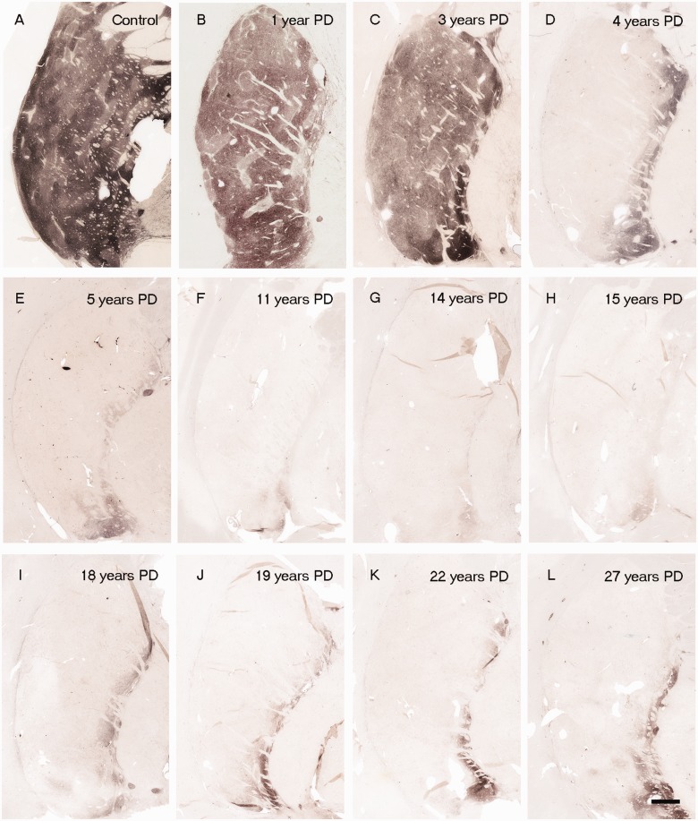
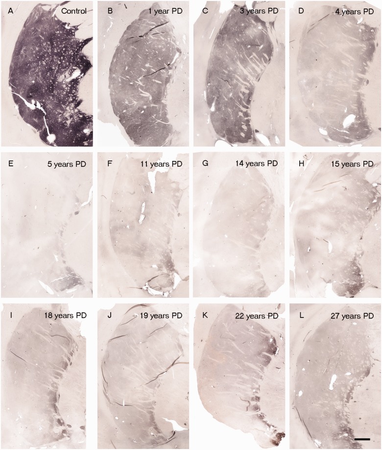
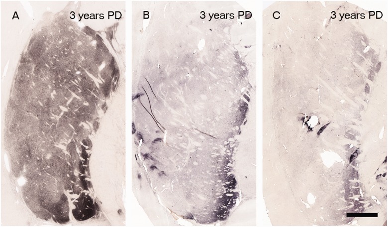
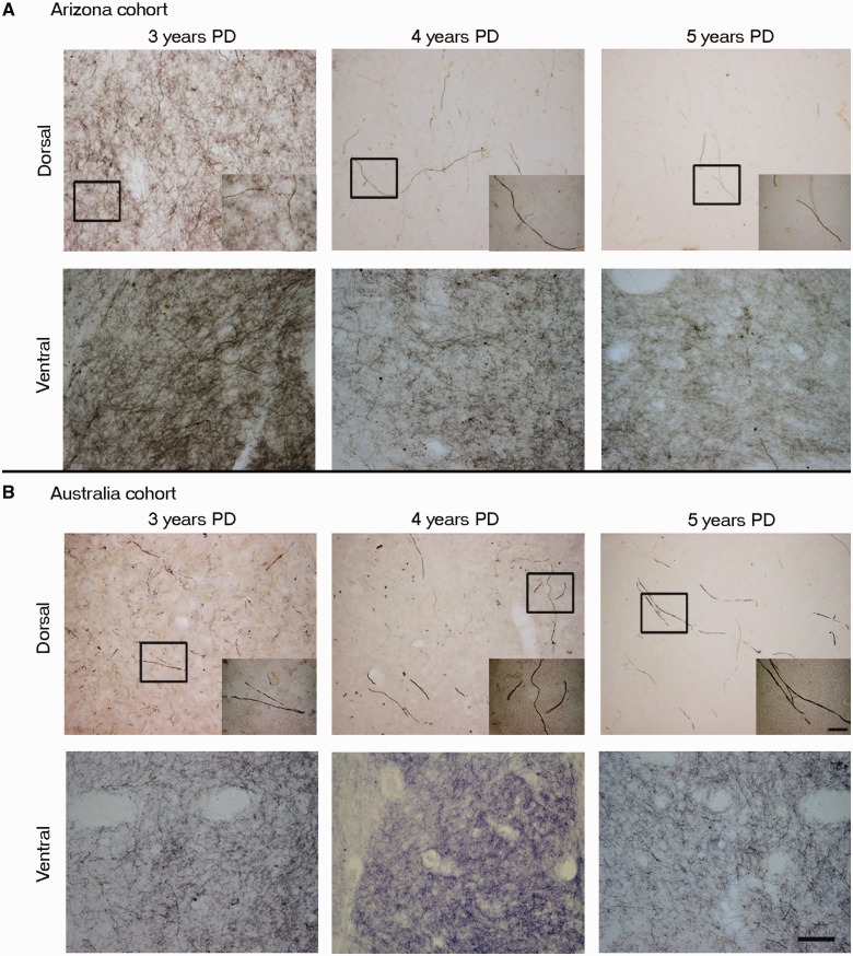
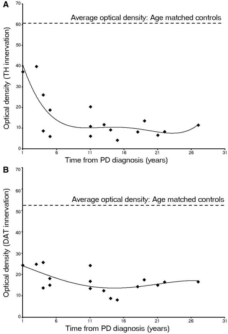
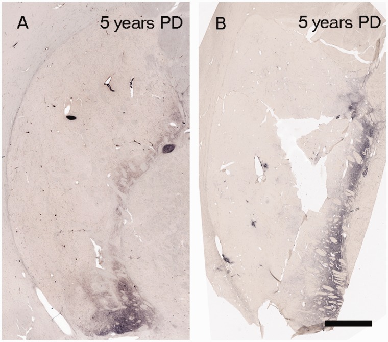
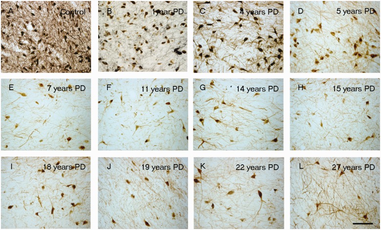
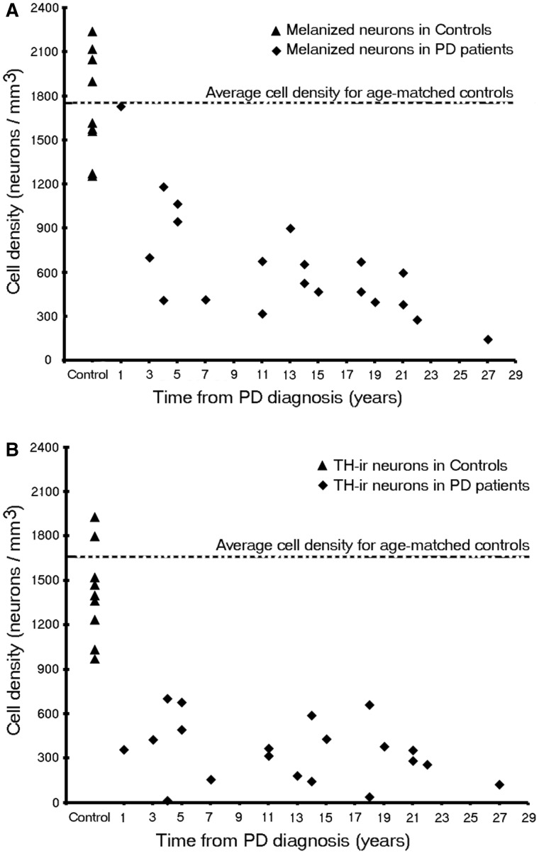
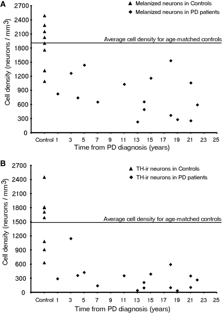
The pace of nigrostriatal degeneration, both with regards to striatal denervation and loss of melanin and tyrosine hydroxylase-positive neurons, is poorly understood especially early in the Parkinson's disease process. This study investigated the extent of nigrostriatal degeneration in patients with Parkinson's disease at different disease durations from time of diagnosis. Brains of patients with Parkinson's disease (n=28) with post-diagnostic intervals of 1-27 years and normal elderly control subjects (n=9) were examined. Sections of the post-commissural putamen and substantia nigra pars compacta were processed for tyrosine hydroxylase and dopamine transporter immunohistochemistry. The post-commissural putamen was selected due to tissue availability and the fact that dopamine loss in this region is associated with motor disability in Parkinson's disease. Quantitative assessments of putaminal dopaminergic fibre density and stereological estimates of the number of melanin-containing and tyrosine hydroxylase-immunoreactive neurons in the substantia nigra pars compacta (both in total and in subregions) were performed by blinded investigators in cases where suitable material was available (n=17). Dopaminergic markers in the dorsal putamen showed a modest loss at 1 year after diagnosis in the single case available for study. There was variable (moderate to marked) loss, at 3 years. At 4 years post-diagnosis and thereafter, there was virtually complete loss of staining in the dorsal putamen with only an occasional abnormal dopaminergic fibre detected. In the substantia nigra pars compacta, there was a 50-90% loss of tyrosine hydroxylase-positive neurons from the earliest time points studied with only marginal additional loss thereafter. There was only a ∼10% loss of melanized neurons in the one case evaluated 1 year post-diagnosis, and variable (30 to 60%) loss during the first several years post-diagnosis with more gradual and subtle loss in the second decade. At all time points, there were more melanin-containing than tyrosine hydroxylase-positive cells. Loss of dopaminergic markers in the dorsal putamen occurs rapidly and is virtually complete by 4 years post-diagnosis. Loss of melanized nigral neurons lags behind the loss of dopamine markers. These findings have important implications for understanding the nature of Parkinson's disease neurodegeneration and for studies of putative neuroprotective/restorative therapies.

Keywords: Parkinsons disease; human brain; morphometry; neuroscience; substantia nigra.


Figures

Figure 1
Figure 1
(A) Tyrosine hydroxylase immunoreactivity in putamen region in control subjects and patients with Parkinson’s disease with varying disease duration from time of diagnosis (n = 28). Representative tyrosine hydroxylase-stained post-commissural putamen sections from a control (A) and patients with Parkinson’s disease, with 1 year (B), 3 years (C), 4 years (D), 5 years (E), 11 years (F), 14 years (G), 15 years (H), 18 years (I), 19 years (J), 22 years (K) and 27 years (L) disease duration. Note the distinct intact tyrosine hydroxylase stained fibre band pattern in the medial putamen. Scale bar in L = 500 µm.
Figure 2
Figure 2
Dopamine transporter immunoreactivity in the putamen region in control subjects and patients with Parkinson’s disease with varying disease duration from time of diagnosis (n = 28). Representative dopamine transporter-stained post-commissural putamen sections from a control (A) and patients with Parkinson’s disease, with 1 year (B), 3 years (C), 4 years (D), 5 years (E), 11 years (F), 14 years (G), 15 years (H), 18 years (I), 19 years (J), 22 years (K) and 27 years (L) disease durations. Scale bar in L = 500 µm.
Figure 3
Figure 3
Tyrosine hydroxylase staining from three cases with disease duration of 3 years following diagnosis: Scale bar in C = 500 µm. PD = Parkinson’s disease.
Figure 4
Figure 4
Comparison between the tyrosine hydroxylase immunoreactivity in the putamen of Arizona versus Australia cases. High magnification photomicrographs of representative dorsal and ventral post-commissural putamen sections comparing Arizona cohort Parkinson’s disease (PD) cases (A) of diseasedurations 3 years, 4 years and 5 years with Australia cohort Parkinson’s disease cases (B) of disease durations 3 years, 4 years and 5 years. Note that the reduction in tyrosine hydroxylase staining at Years 4 and 5 is comparable in the two cohorts, with Year 3 showing more variable results as described in text. Inset demonstrates a single but relatively normal dopamine fibre at Year 3 and isolated morphologically abnormal fibres at Years 4 and 5. Note the relative preservation of the ventral versus dorsal putamen. Scale bars: A and B = 100 µm; insets = 20 µm.
Figure 5
Figure 5
Optical density of putaminal (A) tyrosine hydroxylase and (B) dopamine transporter immunohistochemistry with the progression of Parkinson’s disease (PD; Arizona cohort, n = 17). The data illustrate the modest loss at Year 1–3 after diagnosis, the rapid decline through Years 4–5 and the relatively stable numbers thereafter.
Figure 6
Figure 6
Parkinson’s disease cases 5 years after disease diagnosis from Arizona cohort (82 years) (A) and Australia cohort (53 years) (B).
Figure 7
Figure 7
Tyrosine hydroxylase immunoreactivity (TH-ir) decreases in SN region with the progression of disease. Representative nigra sections from a control (A) and patients with Parkinson’s disease, with a disease duration of 1 year (B), 4 years (C), 5 years (D), 7 years (E), 11 years (F), 14 years (G), 15 years (H), 18 years (I), 19 years (J), 22 years (K) and 27 years (L). Scale bar in L = 100 µm.
Figure 8
Figure 8
Stereologic Estimates of (A) melanin-containing and (B) tyrosine hydroxylase-immunoreactivity (TH-ir) neurons in patients with Parkinson’s disease (PD) of the whole SNc at different disease durations (n = 20).
Figure 9
Figure 9
Stereological estimates of (A) melanin-containing and (B) tyrosine hydroxylase-immunoreactivity (TH-ir) neurons in patients with Parkinson’s disease in the ventrolateral tier of the SNc at different disease durations (n = 16).

References

    1. Bartus RT, Herzog CD, Chu Y, Wilson A, Brown L, Siffert J, et al. Bioactivity of AAV2-neurturin gene therapy (CERE-120): differences between Parkinson's disease and nonhuman primate brains. Mov Disord. 2010;26:27–36. - PMC - PubMed
    1. Beach TG, Adler CH, Sue LI, Peirce JB, Bachalakuri J, Dalsing-Hernandez JE, et al. Reduced striatal tyrosine hydroxylase in incidental Lewy body disease. Acta Neuropathol. 2008a;115:445–51. - PMC - PubMed
    1. Beach TG, Sue LI, Walker DG, Roher AE, Lue L, Vedders L, et al. The Sun Health Research Institute Brain Donation Program: description and experience, 1987-2007. Cell Tissue Bank. 2008b;9:229–45. - PMC - PubMed
    1. Bernheimer H, Birkmayer W, Hornykiewicz O, Jellinger K, Seitelberger F. Brain dopamine and the syndromes of Parkinson and Huntington. Clinical, morphological and neurochemical correlations. J Neurol Sci. 1973;20:415–55. - PubMed
    1. Chu Y, Kompoliti K, Cochran EJ, Mufson EJ, Kordower JH. Age-related decreases in Nurr1 immunoreactivity in the human substantia nigra. J Comp Neurol. 2002;450:203–14. - PubMed

Publication types

Substances