. 2013 Sep;19(9):1226-37.
doi: 10.1261/rna.037077.112. Epub 2013 Jul 24.

Crystal structure of 3WJ core revealing divalent ion-promoted thermostability and assembly of the Phi29 hexameric motor pRNA

Affiliations

Crystal structure of 3WJ core revealing divalent ion-promoted thermostability and assembly of the Phi29 hexameric motor pRNA

Hui Zhang et al. RNA. 2013 Sep.

Abstract

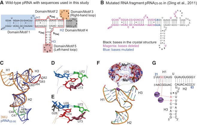
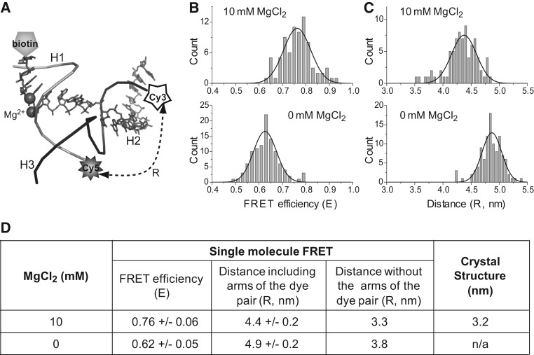
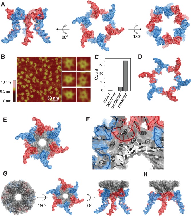
The bacteriophage phi29 DNA packaging motor, one of the strongest biological motors characterized to date, is geared by a packaging RNA (pRNA) ring. When assembled from three RNA fragments, its three-way junction (3WJ) motif is highly thermostable, is resistant to 8 M urea, and remains associated at extremely low concentrations in vitro and in vivo. To elucidate the structural basis for its unusual stability, we solved the crystal structure of this pRNA 3WJ motif at 3.05 Å. The structure revealed two divalent metal ions that coordinate 4 nt of the RNA fragments. Single-molecule fluorescence resonance energy transfer (smFRET) analysis confirmed a structural change of 3WJ upon addition of Mg²⁺. The reported pRNA 3WJ conformation is different from a previously published construct that lacks the metal coordination sites. The phi29 DNA packaging motor contains a dodecameric connector at the vertex of the procapsid, with a central pore for DNA translocation. This portal connector serves as the foothold for pRNA binding to procapsid. Subsequent modeling of a connector/pRNA complex suggests that the pRNA of the phi29 DNA packaging motor exists as a hexameric complex serving as a sheath over the connector. The model of hexameric pRNA on the connector agrees with AFM images of the phi29 pRNA hexamer acquired in air and matches all distance parameters obtained from cross-linking, complementary modification, and chemical modification interference.

Keywords: RNA crystal; RNA nanotechnology; RNA therapeutics; metal ions; three-way junction.


Figures

FIGURE 1.
FIGURE 1.
Secondary structure of phi29 pRNA with the 3WJ core and comparison of the two RNA molecules used in crystallization. (A) Full-length pRNA with the 3WJ motif boxed in red. The three individual RNA strands of the 3WJ are shown as a3wj, b3wj, and c3wj. The helical segments are designated as H1, H2, and H3, respectively. The interlocking loop sequences of 5′aacc in Domain/Motif 3 and 3′uugg in Domain/Motif 5 are indicated by lowercase letters. (B) Sequence of the RNA fragment of the published crystal structure pRNA25-95 (Ding et al. 2011), where the nucleotides deleted are in magenta and the nucleotides mutated are in blue. (C) Superposition of the pRNA25-95 (blue) (Ding et al. 2011) and 3WJ domain (gold) crystal structures. The nucleotides in the left-hand (red) and right-hand (green) loops are also highlighted. (D,E) Comparison of the triple U bulge junction (U72U73U74) for the core junction crystal structures of pRNA25-95 (D) (Ding et al. 2011) and the current 3WJ domain (E). Stereoviews are provided in Supplemental Figure S4. (F) Crystal structure of the pRNA 3WJ with two metal binding sites (magenta). A close-up view of the metal binding sites superposed on the 2Fo−Fc electron density map (blue mesh contoured at 1.0 σ) and the anomalous difference map (red mesh contoured at 4.5 σ) is shown in the inset. (G) Schematic representation of the 3WJ with metal coordinating nucleotides in red. Numbers in blue represent the nucleotide locations in the wild-type pRNA sequence.
FIGURE 2.
FIGURE 2.
Comparison of distances between the 3WJ H2/H3 distal ends derived from FRET measurements (with and without 10 mM MgCl2) and crystal data. (A) Location of labeling in 3WJ for single-molecule FRET study. (a3wj indicated by gray sticks; b3wj, black backbone; c3wj, gray backbone.) (B,C) Comparison of the FRET efficiency (B) and distance (C) at 0 mM and 10 mM Mg2+, respectively. The Gaussian curve fittings used to locate the peak of the distributions are shown as black lines. (D) Comparison of the distances from FRET and the crystal structure. The distance was corrected by subtracting the arm sizes of the fluorophore pair (Shu et al. 2010).
FIGURE 3.
FIGURE 3.
Effect of magnesium ions on the resistance of pRNA 3WJ to urea denaturation, assayed by 15% PAGE gels with (A) 2 mM EDTA and (B) 5 mM MgCl2. (Lane 1) Single RNA oligo b3wj; (lane 2) sample annealed from two oligos a3wj and b3wj; (lane 3) 3WJ assembled from three oligos a3wj, b3wj, and c3wj; and (lane 4) DNA ladder.
FIGURE 4.
FIGURE 4.
Structure of a pRNA hexamer ring and docking of the hexameric pRNA model with the dodecameric connector ring of phi29. (A) A 3D model of hexameric pRNA based on the crystal structure of 3WJ with views from three mutually perpendicular angles. (B) AFM images of hexameric re-engineered pRNA rings show strong correlation in size and shape with the 3D computer models for the intact, full-length pRNA hexamers. (C) Histogram of RNA particles in AFM images with discernible stoichiometry. (D) Top view of the published 3D computer model of hexameric pRNA constructed based on biochemical data (PDB 1L4O) (Hoeprich and Guo 2002). (E) Model of a pRNA hexamer complexed with a dodecameric connector ring based on the 3WJ and connector (PDB 1H5W) (Guasch et al. 2002) crystal structures. (For an animation movie of the phi29 pRNA-connector model, see Supporting Video S1.) (F) A close-up view of proper anchoring of the connector N-terminal helices for optimal interactions with pRNA. (G) Three mutually perpendicular views of the hexameric pRNA-connector assembly. (H) A side-view of the 3D model of the hexameric pRNA-connector assembly based on biochemical data (PDB 1L4P) (Hoeprich and Guo 2002).
FIGURE 5.
FIGURE 5.
Confirmation of the close proximity between two nucleotide groups (black and gray) in the pRNA model based on the 3WJ crystal structure, in agreement with the published data revealed by (A) complementary modification; (B) psoralen crosslinking; (C) Phenphi crosslinking; and (DF) azidophenacyl crosslinking.

References

    1. Abdelmawla S, Guo S, Zhang L, Pulukuri S, Patankar P, Conley P, Trebley J, Guo P, Li QX 2011. Pharmacological characterization of chemically synthesized monomeric pRNA nanoparticles for systemic delivery. Mol Ther 19: 1312–1322 - PMC - PubMed
    1. Adams PD, Grosse-Kunstleve RW, Hung LW, Ioerger TR, McCoy AJ, Moriarty NW, Read RJ, Sacchettini JC, Sauter NK, Terwilliger TC 2002. PHENIX: Building new software for automated crystallographic structure determination. Acta Crystallogr D Biol Crystallogr 58: 1948–1954 - PubMed
    1. Adams PD, Afonine PV, Bunkoczi G, Chen VB, Davis IW, Echols N, Headd JJ, Hung LW, Kapral GJ, Grosse-Kunstleve RW, et al. 2010. PHENIX: A comprehensive Python-based system for macromolecular structure solution. Acta Crystallogr D Biol Crystallogr 66: 213–221 - PMC - PubMed
    1. Afonin KA, Bindewald E, Yaghoubian AJ, Voss N, Jacovetty E, Shapiro BA, Jaeger L 2010. In vitro assembly of cubic RNA-based scaffolds designed in silico. Nat Nanotechnol 5: 676–682 - PMC - PubMed
    1. Al-Hashimi HM, Walter NG 2008. RNA dynamics: It is about time. Curr Opin Struct Biol 18: 321–329 - PMC - PubMed

Publication types