. 2013 Oct;87(19):10612-27.
doi: 10.1128/JVI.01370-13. Epub 2013 Jul 24.

Morphological and biochemical characterization of the membranous hepatitis C virus replication compartment

Affiliations

Morphological and biochemical characterization of the membranous hepatitis C virus replication compartment

David Paul et al. J Virol. 2013 Oct.

Abstract

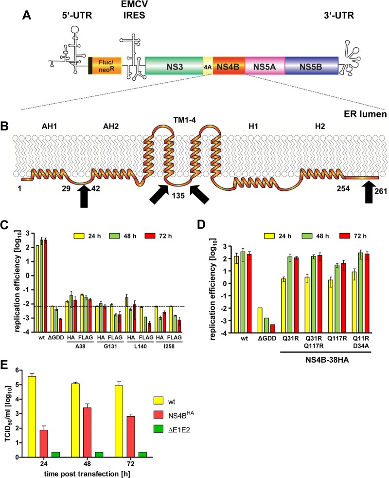
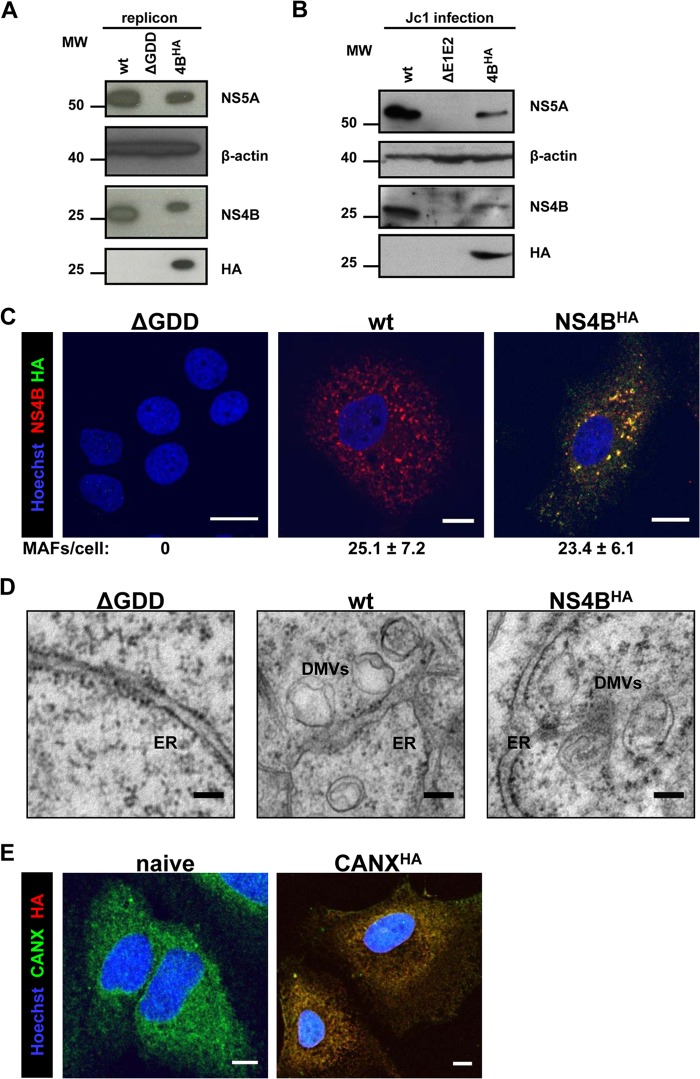
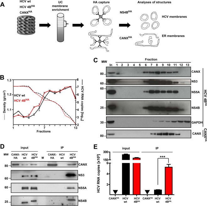
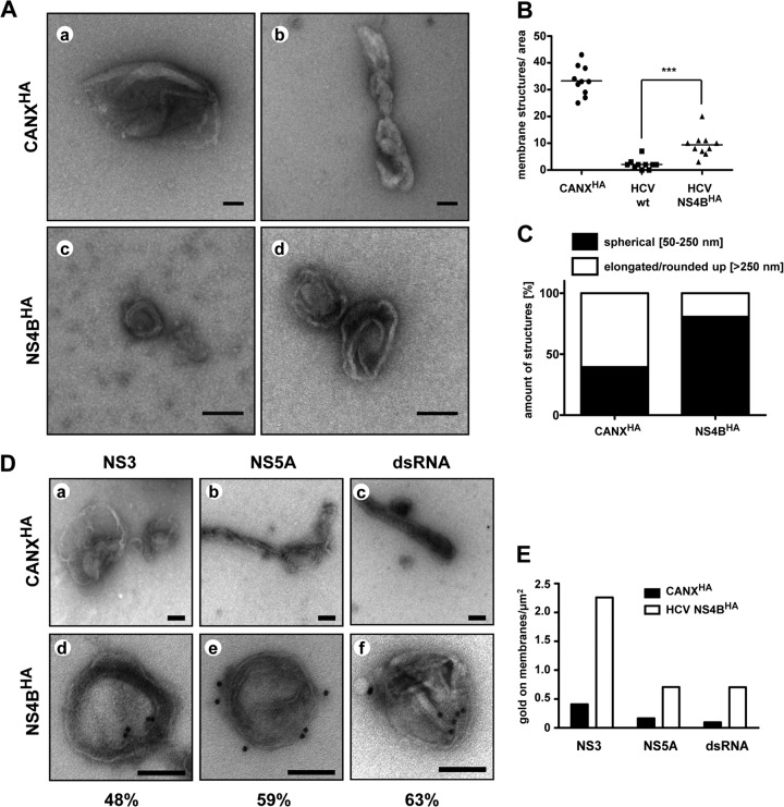
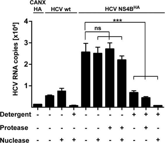
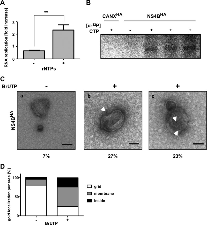
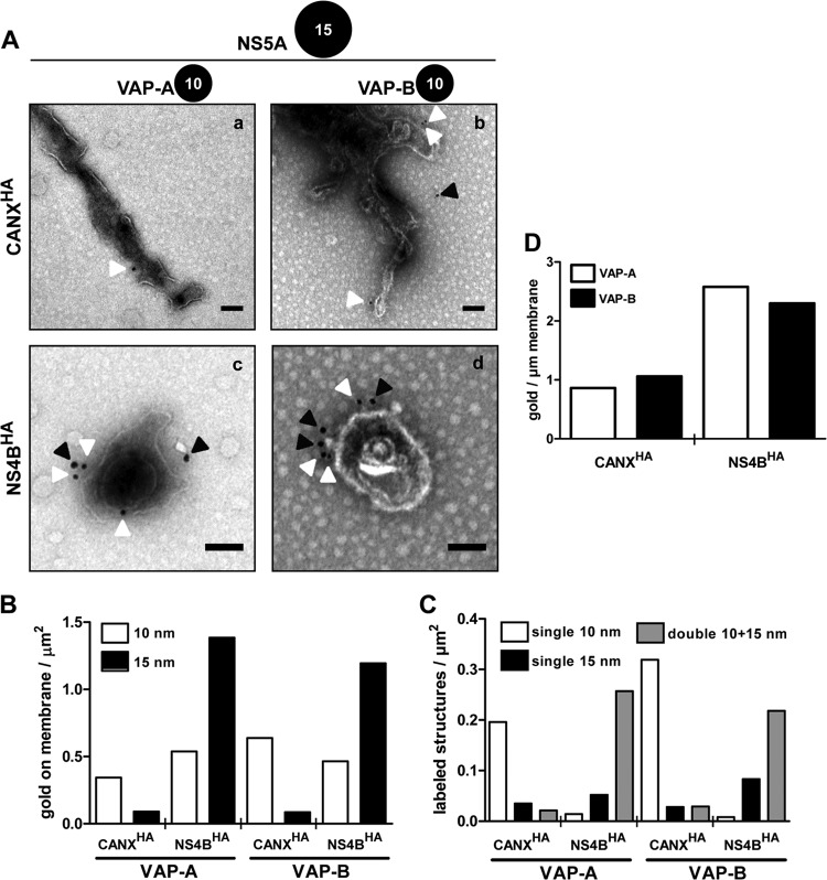
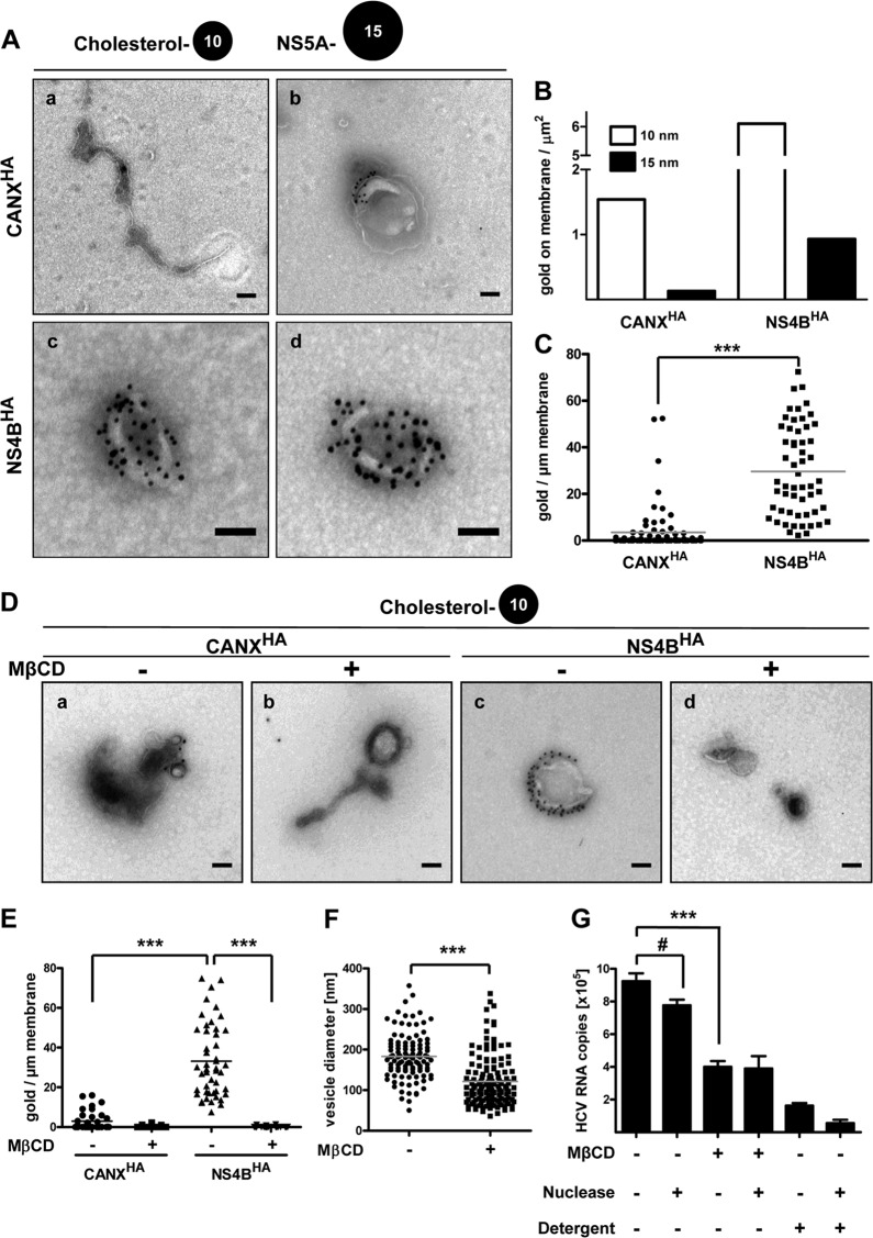
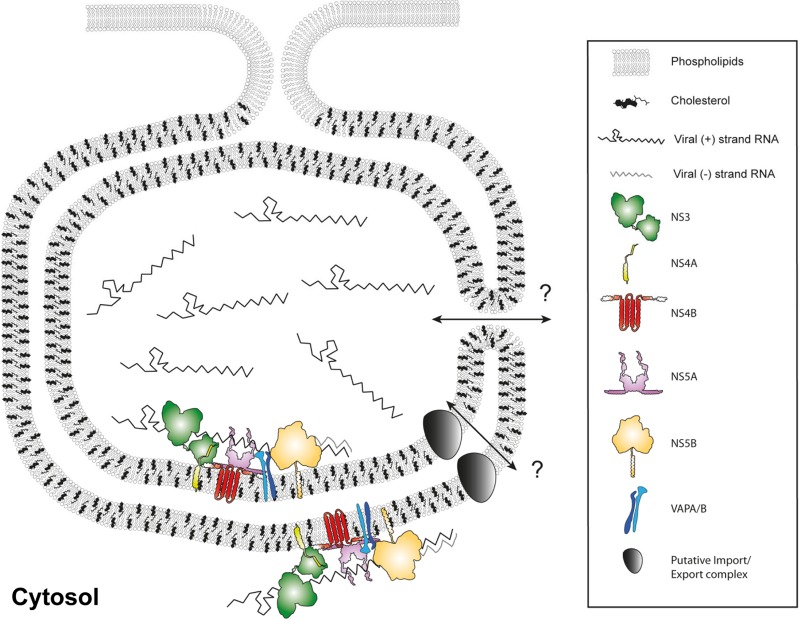
Like all other positive-strand RNA viruses, hepatitis C virus (HCV) induces rearrangements of intracellular membranes that are thought to serve as a scaffold for the assembly of the viral replicase machinery. The most prominent membranous structures present in HCV-infected cells are double-membrane vesicles (DMVs). However, their composition and role in the HCV replication cycle are poorly understood. To gain further insights into the biochemcial properties of HCV-induced membrane alterations, we generated a functional replicon containing a hemagglutinin (HA) affinity tag in nonstructural protein 4B (NS4B), the supposed scaffold protein of the viral replication complex. By using HA-specific affinity purification we isolated NS4B-containing membranes from stable replicon cells. Complementing biochemical and electron microscopy analyses of purified membranes revealed predominantly DMVs, which contained viral proteins NS3 and NS5A as well as enzymatically active viral replicase capable of de novo synthesis of HCV RNA. In addition to viral factors, co-opted cellular proteins, such as vesicle-associated membrane protein-associated protein A (VAP-A) and VAP-B, that are crucial for viral RNA replication, as well as cholesterol, a major structural lipid of detergent-resistant membranes, are highly enriched in DMVs. Here we describe the first isolation and biochemical characterization of HCV-induced DMVs. The results obtained underline their central role in the HCV replication cycle and suggest that DMVs are sites of viral RNA replication. The experimental approach described here is a powerful tool to more precisely define the molecular composition of membranous replication factories induced by other positive-strand RNA viruses, such as picorna-, arteri- and coronaviruses.


Figures

Fig 1
Fig 1
HCV tolerates an HA tag insertion into the N-terminal region of NS4B. (A) Schematic representation of the bicistronic HCV subgenomic reporter replicon. Firefly luciferase (Fluc) or neomycin-phosphotransferase (neoR) is expressed as an N-terminal fusion with 16 amino acids of the N-terminal region of the core protein (black line) and is translated under the control of the HCV IRES contained in the 5′ UTR. The second cistron (NS3 to NS5B) is translated via the IRES of the encephalomyocarditis virus (EMCV-IRES). (B) Predicted NS4B membrane topology. N-terminal amphipathic α-helices AH1 and AH2, the four transmembrane segments (TM1-4), and the C-terminal α-helices H1 and H2 are schematically depicted. Numbers indicate amino acid positions of the JFH1 isolate. Affinity-tag insertion sites are highlighted by black arrows. (C) Huh7-Lunet cells were transfected with the in vitro-transcribed luciferase replicon RNAs specified at the bottom. Cells were lysed 4, 24, 48, and 72 h after transfection, and the luciferase activity in cell lysates was determined. Data were normalized to the 4-h value that reflects transfection efficiency. The background of the assay is determined by the NS5B active-site polymerase mutant (ΔGDD) (dashed line). Mean values of two independent experiments are shown. Error bars indicate standard deviations. (D) Huh7-Lunet cells were transfected with the in vitro-transcribed luciferase replicon RNAs specified at the bottom. Replication efficiency was determined as described for panel C. (E) Release kinetics of infectious HCV particles. Huh7-Lunet cells were transfected with the full-length HCV RNAs specified on the right. Culture supernatants were harvested at the given time points. Infectivity titers were determined by limiting-dilution assay and are expressed as 50% tissue culture infective doses (TCID50)/ml. Mean values of two independent experiments are shown; error bars indicate standard deviations.
Fig 2
Fig 2
Characterization of HA-tagged NS4B. (A) Huh7-Lunet cells were transfected with the in vitro-transcribed luciferase replicon RNAs specified at the top. Cells were harvested 72 h posttransfection and analyzed by immunoblotting, using the monospecific antibodies indicated on the right. The positions of molecular weight (MW; in thousands) markers are depicted on the left. (B) Huh7.5 cells were infected with the culture supernatants of cells transfected with the Jc1-derived full-length constructs specified at the top. After 72 h, cells were harvested and processed as described for panel A. (C) Huh7-Lunet cells were transfected with the in vitro-transcribed luciferase replicon RNAs specified at the top of each panel. After 48 h, cells were fixed, permeabilized with digitonin, and stained with NS4B- and HA-specific antibodies prior to confocal immunofluorescence microscopy. Only merged images are shown. Scale bars represent 5 μm. Numbers below each panel indicate the mean ± the standard deviation (SD) for NS4B-containing MAF per cell; for each condition, at least 10 different HCV-positive cells were analyzed. (D) Huh7-Lunet cells were transfected with in vitro-transcribed luciferase replicon RNAs as specified at the top of each panel. After 48 h, cells were fixed and flat embedded for TEM analysis. Scale bars represent 100 nm. (E) Naive Huh7-Lunet cells and those overexpressing CANXHA were fixed, permeabilized with Triton X-100, and stained with CANX- and HA-specific antibodies prior to confocal immunofluorescence microscopy. Only merged images are shown. Scale bars represent 5 μm.
Fig 3
Fig 3
Purification and biochemical characterization of NS4BHA-associated membranes. (A) Schematic overview of HCV membrane preparation. Cells containing a stably replicating wild-type replicon (HCV wt), the replicon sg4BHA31R (HCV 4BHA), and control cells stably overexpressing CANXHA were broken by hypotonic lysis. Postnuclear cytosolic supernatants were separated by discontinuous sucrose gradient ultracentrifugation (UC). Subsequently, membrane fractions were pooled and subjected to affinity capture using HA beads. After elution with the HA peptide, purified material was used for further analyses. (B) Distribution of HCV RNA (right y axis, solid lines) and corresponding density of each fraction (left y axis, dashed lines) along the gradient. Mean values and error bars indicating the standard deviations of three independent measurements are depicted. (C) Fractions analyzed for their protein content are shown for HCV NS4BHA. Proteins were separated by SDS-PAGE, and monospecific antibodies were used to detect calnexin (CANX), glyceraldehyde-3-phosphate dehydrogenase (GAPDH), NS3, NS5A, and NS4B, as indicated on the right. The positions of molecular weight (MW) markers are depicted on the left. (D) Protein content after HA-specific affinity capture. Samples were analyzed as described for panel C. (E) HCV RNA content in purified material. Mean values with standard deviations, as indicated by the error bars, from at least three independent experiments, with three measurements each, are given. No HCV RNA was detected in CANXHA samples, as indicated by the inverted triangles (▼). ***, P < 0.0001.
Fig 4
Fig 4
Morphological characterization of purified HCV-remodeled membranes. (A) HA-captured material was negatively stained with uranylacetate and examined by TEM. Representative membranous structures from NS4BHA- and control CANXHA-captured material are shown. Scale bars represent 100 nm. (B) Number of membrane structures per area of 10 randomly chosen hexagons (area/hexagon, ∼2,913 μm2) are given. Horizontal lines indicate mean values. ***, P < 0.0005. (C) Quantification of membrane morphotypes. More than 100 structures per sample were classified as specified at the top and are represented as relative values. (D) HA affinity-purified membranes of CANXHA (a through c) and HCV NS4BHA cells (d through f) were immunolabeled with antibodies recognizing NS3 (a and d), NS5A (b and e), and dsRNA (c and f). Samples were negatively stained with uranylacetate and examined by TEM. The scale bar in each panel corresponds to 100 nm. Percentages of labeled DMVs (n > 75) are given below panels d through f. (E) Quantification of immunolabeling. At least 100 gold particles were counted, and fractions of gold particles associated with membranes per area of the grid are depicted.
Fig 5
Fig 5
HCV RNA associated with affinity-purified membranes is protected against nuclease. Equal aliquots of NS4BHA-purified and unspecifically bound (NS4B wt) material were subjected to treatment with nuclease (1 U/μl Benzonase), protease (8 mg/ml proteinase K), and detergent (1% Triton X-100) for 1 h at 25°C as indicated at the bottom. Subsequently, HCV RNA was extracted and quantified by quantitative reverse transcription-PCR. Mean values and error bars indicating the standard deviations for at least three independent experiments are depicted. ns, P > 0.4; ***, P < 0.0003.
Fig 6
Fig 6
HCV-induced double-membrane vesicles are sites of RNA replication. (A) Equal fractions of NS4BHA-purified material were incubated without (−) or with (+) exogenously added ribonucleotides in an in vitro replicase assay as specified at the bottom. Total HCV RNA was quantified before and after the in vitro replicase assay and is represented as the fold increase. Mean values and error bars representing standard deviations from six replicates in two independent experiments are shown. **, P < 0.001. (B) Equal amounts of control CANXHA and NS4BHA-purified membranes were subjected to an in vitro replicase assay in the absence (−) or presence (+) of radioactively labeled [α-32P]CTP. After RNA purification, samples were analyzed by denaturing glyoxal agarose gel electrophoresis and autoradiography. (C) Membranes from the NS4BHA purification were used for in vitro replicase assay in the absence (−) or presence (+) of BrUTP. After immunolabeling with a BrdU-specific antibody and subsequent negative staining, samples were examined by TEM. White arrowheads show the locations of gold particles. The number below each panel indicates the percentage of non-specifically gold-labeled DMVs (a) or of DMVs with gold labeling on the exterior (b) or apparent in the interior (c) (n > 75). The scale bar in each panel corresponds to 100 nm. (D) Quantification of immunolabeling. At least 100 gold particles were counted, and the fraction of gold particles with respect to their location per area is depicted.
Fig 7
Fig 7
HCV–co-opted host proteins VAP-A and VAP-B are enriched in DMVs. (A) Purified ER control (a and b) and HCV-remodeled membranes (c and d) were consecutively labeled for NS5A (15-nm gold) and for VAP-A (a and c) or VAP-B (b and d) (10-nm gold). After negative staining, they were examined by TEM. White and black arrowheads highlight 10- and 15-nm gold particles, respectively. Scale bars represent 100 nm. (B) Quantification of gold particles on membranes. At least 100 gold particles were counted, and the number of gold particles associated with membranes per area of the grid is depicted. (C) Number of single- and double-labeled structures containing either NS5A or VAP-A/B, or both viral and cellular protein, respectively, per area on the grid. At least 50 membranous structures per condition were analyzed. (D) Amount of 10-nm colloidal gold particles per micrometer of membrane length. Only membranes positive for NS5A were considered for the analysis of NS4BHA-captured membranes. The analysis is based on ∼50 membranous structures with a total membrane length of more than 50 μm for each condition.
Fig 8
Fig 8
DMV membranes contain large amounts of cholesterol. (A) Purified ER control (a and b) and HCV-remodeled membranes (c and d) were consecutively labeled for cholesterol using biotinylated perfringolysin O (10-nm gold) and for NS5A (15-nm gold). After negative staining, they were examined by TEM. Scale bars correspond to 100 nm. (B) Quantification of gold particles on membranes. At least 100 gold particles were counted, and the number of gold particles associated with membranes per area of the grid is depicted. (C) Amount of 10-nm colloidal gold particles per micrometer of membrane length of a given structure. Only membranes also positive for NS5A were considered for the analysis of NS4BHA-captured membranes. The analysis is based on at least 75 membranous structures with a total membrane length of more than 60 μm for each condition. Horizontal lines indicate mean values. ***, P < 0.0001. (D) Purified ER control (CANXHA) and HCV-remodeled membranes (NS4BHA) were treated with 10 mM MβCD for 1 h at 25°C as indicated, subsequently labeled for cholesterol using biotinylated perfringolysin O, and after negative staining examined by TEM. Scale bars correspond to 100 nm. (E) Amount of 10-nm colloidal gold particles per micrometer of membrane length of a given structure. The analysis is based on at least 50 membranous structures with a total membrane length of more than 50 μm for each condition. Horizontal lines indicate mean values. ***, P < 0.0001. (F) DMV diameters after MβCD treatment. Membranes from the NS4BHA capture were MβCD treated as described for panel D, and DMV diameters were measured. Horizontal lines indicate mean values. n > 100; ***, P < 0.0001. (G) Equal aliquots of NS4BHA-purified material were subjected to treatment with methyl-β-cyclodextrin (MβCD; 10 mM), nuclease (1 U/μl Benzonase), and/or detergent (1% Triton X-100) for 1 h at 25°C as indicated at the bottom. HCV RNA was extracted and quantified by reverse transcription-quantitative PCR. Mean values and error bars indicating the standard deviations for three independent experiments are depicted. #, P > 0.05; ***, P < 0.0001.
Fig 9
Fig 9
Schematic model of an HCV-induced DMV. Cellular lipids, viral RNA, and viral and cellular proteins are depicted as indicated in the panel on the right. The HCV replicase complex is associated with DMV membranes, and the bulk of viral RNA resides in a nuclease-protected environment, likely the DMV lumen. HCV creates a unique membrane environment containing co-opted cellular proteins, exemplified here by VAP-A/B, and also modifies the membrane lipid composition, highlighted by the large quantity of cholesterol. It remains to be determined how transport to and out of the DMV interior might be mediated, as indicated by the question marks. Possible scenarios are not (yet) completely sealed DMVs or a distinct transporter function.

References

    1. Lavanchy D. 2009. The global burden of hepatitis C. Liver Int. 29(Suppl 1):74–81 - PubMed
    1. Sarrazin C, Hezode C, Zeuzem S, Pawlotsky JM. 2012. Antiviral strategies in hepatitis C virus infection. J. Hepatol. 56(Suppl 1):S88–S100 - PubMed
    1. Vermehren J, Susser S, Lange CM, Forestier N, Karey U, Hughes E, Ralston R, Tong X, Zeuzem S, Sarrazin C. 2012. Mutations selected in the hepatitis C virus NS3 protease domain during sequential treatment with boceprevir with and without pegylated interferon alfa-2b. J. Viral Hepat. 19:120–127 - PubMed
    1. van Regenmortel MHV, Fauquet CM, Bishop DHL, Carstens EB, Estes MK, Lemon SM, Maniloff J, Mayo MA, McGeoch DJ, Pringle CR, Wickner RB. (ed). 2000. Virus taxonomy. Seventh report of the International Committee on Taxonomy of Viruses. Academic Press, San Diego, CA
    1. Simmonds P, Bukh J, Combet C, Deleage G, Enomoto N, Feinstone S, Halfon P, Inchauspe G, Kuiken C, Maertens G, Mizokami M, Murphy DG, Okamoto H, Pawlotsky JM, Penin F, Sablon E, Shin I, Stuyver LJ, Thiel HJ, Viazov S, Weiner AJ, Widell A. 2005. Consensus proposals for a unified system of nomenclature of hepatitis C virus genotypes. Hepatology 42:962–973 - PubMed

Publication types

MeSH terms