. 2013 Oct;87(19):10660-7.
doi: 10.1128/JVI.01539-13. Epub 2013 Jul 24.

Circulating dendritic cells isolated from healthy seropositive donors are sites of human cytomegalovirus reactivation in vivo

Affiliations

Circulating dendritic cells isolated from healthy seropositive donors are sites of human cytomegalovirus reactivation in vivo

Matthew B Reeves et al. J Virol. 2013 Oct.

Abstract

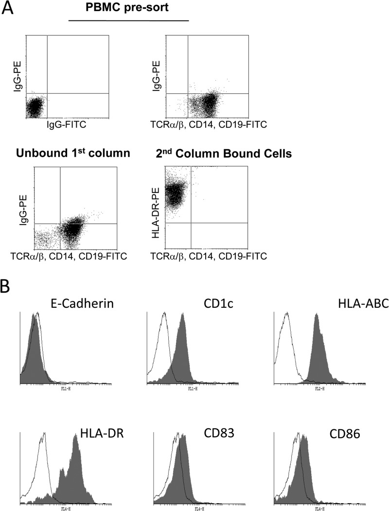
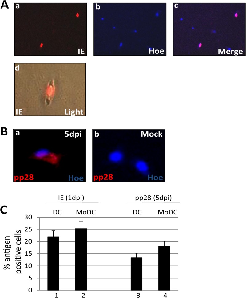
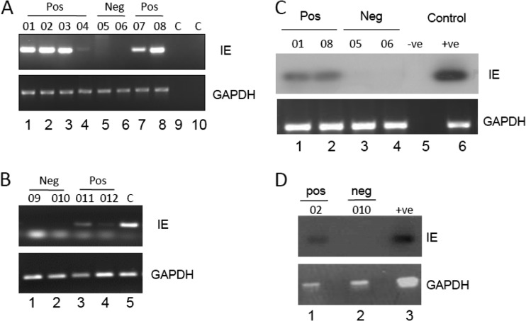
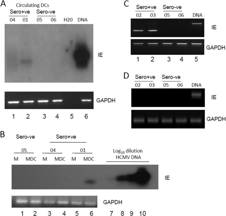
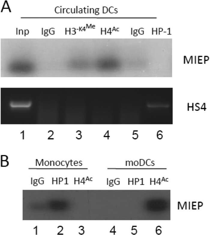
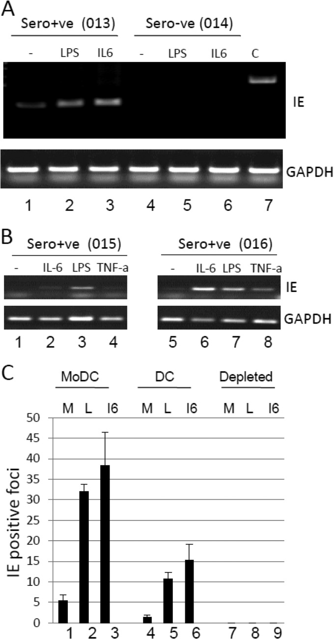
Primary infection with human cytomegalovirus (HCMV) is generally asymptomatic in healthy individuals and results in a lifelong infection of the host. In contrast, in immunosuppressed transplant recipients and late-stage AIDS patients, HCMV infection and reactivation can result in severe disease or death. In vivo, latency is established in bone marrow CD34(+) progenitor cells with reactivation linked with their differentiation to macrophages and dendritic cells (DCs). However, previous analyses have relied on ex vivo differentiation of myeloid progenitor cells to DCs in culture. Here, we now report on the isolation and analysis of circulating blood myeloid DCs, resulting from natural differentiation in vivo, from healthy HCMV-seropositive carriers. We show that these in vivo-differentiated circulating DCs are fully permissive for HCMV and exhibit a phenotype similar to that of monocyte-derived DCs routinely used for in vitro studies of HCMV. Importantly, we also show that these DCs from healthy HCMV-seropositive donors carry HCMV genomes and, significantly, are typically positive for viral immediate-early (IE) gene expression, in contrast to circulating monocytes, which carry genomes with an absence of IE expression. Finally, we show that HCMV reactivation from these circulating DCs is enhanced by inflammatory stimuli. Overall, these data argue that the differentiation in vivo of myeloid progenitors to circulating DCs promotes the reactivation of HCMV lytic gene expression in healthy individuals, thereby providing valuable confirmation of studies performed using in vitro generation of DCs from myeloid precursors to study HCMV reactivation.


Figures

Fig 1
Fig 1
Circulating myeloid dendritic cells can be isolated from peripheral blood. (A) Flow cytometric analysis was performed on peripheral blood mononuclear cells stained with FITC-conjugated anti-TCRα/β, -CD14, and -CD19 antibodies (top right) or isotype-matched antibody controls (top left). Following magnetic column depletion with anti-CD3, -CD7, -CD16, -CD56, and -CD123 antibodies, the unbound fraction was stained with FITC-conjugated anti-TCRα/β, -CD14, and -CD19 antibodies (bottom). CD4-positive cells were depleted from the unbound fraction and stained with FITC-conjugated anti-TCRα/β, -CD14, and -CD19 antibodies and a PE-conjugated anti-HLA-DR antibody. (B) Isolated DCs were stained with a panel of cell surface markers (filled histogram) or an isotype-matched control (open histogram) immediately postisolation.
Fig 2
Fig 2
Directly isolated DCs are permissive for HCMV gene expression. (A) Three hours postisolation, DCs were infected with a myelotropic stock of TB40/e and stained for IE gene (a) and nuclear DNA (b) 24 h postinfection. The merged image (c) shows nuclear IE gene expression. DC morphology after isolation and infection is shown in an IE gene-positive cell shown merged with bright-field image (d). (B) TB40/e-infected (a) or mock-infected (b) DCs were costained for pp28 expression and nuclear DNA 5 days postinfection. (C) DCs and monocyte-derived DCs infected with TB40/e were stained for IE (bars 1 and 2) or pp28 (bars 3 and 4) expression, and the number of antigen-positive cells was determined. Data show enumeration from 10 fields of view performed in triplicate. dpi, days postinfection.
Fig 3
Fig 3
Detection of HCMV DNA sequences in circulating blood DCs of seropositive donors. (A and B) Nested PCR analysis of DCs from multiple seropositive (donors 01 to 04, 07, 08, 011, and 012), seronegative (donors 05, 06, 09, and 010), or water (panel A, lanes 9 and 10) and DNA (panel B, lane 5) controls was performed using IE gene-specific (a) and glyceraldehyde-3-phosphate dehydrogenase (GAPDH) DNA-specific (b) PCRs. Nested PCR gave rise to a 312-bp product from HCMV DNA. (C and D) A single-round IE gene PCR analysis on three seropositive (donors 01, 02, and 08), three seronegative donors (05, 06, and 010), and a water (panel C, lane 5) or DNA (panel C, lane 6, and panel D, lane 3) control was analyzed by Southern blotting and probing with an IE gene-specific probe that detected a 310-bp product. A GAPDH PCR was used to confirm the isolation of DNA.
Fig 4
Fig 4
Detection of IE RNA transcripts in circulating blood DCs of seropositive donors. (A) RNA prepared from DCs directly isolated from the peripheral blood of seropositive (donors 04 and 01) and seronegative (donors 05 and 06) donors was amplified in an IE gene RT-PCR. Water or HCMV DNA was amplified as a control for the viral PCRs (lane 5 and 6). Amplified products were then analyzed by Southern blotting with an IE gene-specific probe. (B) Monocytes isolated from donors 04, 01, and 05 were analyzed either directly postisolation (lanes 1, 3, and 5) or after differentiation to MoDCs (lanes 2, 4, and 6) for IE RNA or GAPDH expression by Southern blotting. A log dilution of viral DNA was used as a positive control (lanes 7 to 10). (C and D) RNA extracted from DCs (C) or monocytes (D) isolated from healthy seropositive (donors 02 and 03) or seronegative (donors 05 and 06) donors was analyzed using a nested intron-spanning RT-PCR for IE72 gene expression. Viral DNA was amplified as a control (lane 5). GAPDH controls are shown (lanes 1 to 5).
Fig 5
Fig 5
The MIEP in circulating DCs is predominantly associated with markers of transcriptional activation. (A) Chromatin immunoprecipitation assays were performed on seropositive DCs with rabbit IgG (lane 2), anti-dimethylated histone H3-K4 (lane 3), anti-acetylated H4 (lane 4), anti-rat IgG (lane 5), or anti-HP1β (lane 6) antibodies. DNA amplified in an MIEP-specific PCR was then analyzed by Southern blotting using an MIEP-specific probe. Alternatively, samples were amplified using primers against the HS4 region of the gamma globulin gene. (B) Monocytes isolated from the same donor were either subjected to ChIP (lanes 1 to 3) or differentiated to MoDCs and then subjected to ChIP analysis (lanes 4 to 6). Immunoprecipitation with IgG (lanes 1 and 3), anti-HP-1 (lanes 2 and 5) or anti-acetylated histone H4 (lanes 3 and 6) was performed, and samples were amplified in an MIEP PCR and analyzed by Southern blotting.
Fig 6
Fig 6
Inflammatory stimulation of DCs enhances reactivation of HCMV. (A) Isolated DCs from a seropositive (donor 013) or seronegative (donor 014) donor were cultured alone (lane 1) or with LPS (lane 2) or IL-6 (lane 3) and analyzed for IE72 and GAPDH RNA expression 16 h poststimulation. (B) Isolated DCs from two further seropositive donors (donors 015 and 016) were left unstimulated (lanes 1 and 5) or stimulated with IL-6 (lanes 2 and 6), LPS (lanes 3 and 7), or TNF-α (lanes 4 and 8) and analyzed for IE72 or GAPDH RNA expression 16 h poststimulation. (C) To test for viral reactivation, 5 × 10⁵ MoDCs (lanes 1 to 3), directly isolated DCs (lanes 4 to 6), or DC- and monocyte-depleted cells (lanes 7 to 9) were cultured alone (M) or with LPS (L) or IL-6 (I6) and then cocultured with fibroblasts for 21 days. The fibroblast monolayer was then stained for IE gene expression and the average number of IE gene-positive foci scored from 5 fields of view.

References

    1. Limaye AP, Kirby KA, Rubenfeld GD, Leisenring WM, Bulger EM, Neff MJ, Gibran NS, Huang ML, Santo Hayes TK, Corey L, Boeckh M. 2008. Cytomegalovirus reactivation in critically ill immunocompetent patients. JAMA 300:413–422 - PMC - PubMed
    1. Peggs KS, Mackinnon S. 2004. Cytomegalovirus: the role of CMV post-haematopoietic stem cell transplantation. Int. J. Biochem. Cell Biol. 36:695–701 - PubMed
    1. Revello MG, Gerna G. 2004. Pathogenesis and prenatal diagnosis of human cytomegalovirus infection. J. Clin. Virol. 29:71–83 - PubMed
    1. Goodrum FD, Jordan CT, High K, Shenk T. 2002. Human cytomegalovirus gene expression during infection of primary hematopoietic progenitor cells: a model for latency. Proc. Natl. Acad. Sci. U. S. A. 99:16255–16260 - PMC - PubMed
    1. Hahn G, Jores R, Mocarski ES. 1998. Cytomegalovirus remains latent in a common precursor of dendritic and myeloid cells. Proc. Natl. Acad. Sci. U. S. A. 95:3937–3942 - PMC - PubMed

Publication types

MeSH terms