. 2013 Oct;87(19):10447-59.
doi: 10.1128/JVI.00049-13. Epub 2013 Jul 24.

Neutralizing polyclonal IgG present during acute infection prevents rapid disease onset in simian-human immunodeficiency virus SHIVSF162P3-infected infant rhesus macaques

Affiliations

Neutralizing polyclonal IgG present during acute infection prevents rapid disease onset in simian-human immunodeficiency virus SHIVSF162P3-infected infant rhesus macaques

J Pablo Jaworski et al. J Virol. 2013 Oct.

Abstract

Simian-human immunodeficiency virus (SHIV) models for human immunodeficiency virus (HIV) infection have been widely used in passive studies with HIV neutralizing antibodies (NAbs) to test for protection against infection. However, because SHIV-infected adult macaques often rapidly control plasma viremia and any resulting pathogenesis is minor, the model has been unsuitable for studying the impact of antibodies on pathogenesis in infected animals. We found that SHIVSF162P3 infection in 1-month-old rhesus macaques not only results in high persistent plasma viremia but also leads to very rapid disease progression within 12 to 16 weeks. In this model, passive transfer of high doses of neutralizing IgG (SHIVIG) prevents infection. Here, we show that at lower doses, SHIVIG reduces both plasma and peripheral blood mononuclear cell (PBMC)-associated viremia and mitigates pathogenesis in infected animals. Moreover, production of endogenous NAbs correlated with lower set-point viremia and 100% survival of infected animals. New SHIV models are needed to investigate whether passively transferred antibodies or antibodies elicited by vaccination that fall short of providing sterilizing immunity impact disease progression or influence immune responses. The 1-month-old rhesus macaque SHIV model of infection provides a new tool to investigate the effects of antibodies on viral replication and clearance, mechanisms of B cell maintenance, and the induction of adaptive immunity in disease progression.


Figures

Fig 1
Fig 1
Effect of passively transferred IgG on plasma viral load in SHIVSF162P3-infected 1-month-old rhesus macaques. Blood samples were collected at regular intervals after viral exposure, the RNA was isolated from plasma, and viral RNA (vRNA) was quantified by real-time RT-PCR. Curves represent individual infected animals that were treated with normal IgG only (n = 6) (A), normal IgG plus MAb b12 (n = 6) (B), SHIVIG only (n = 6) (C), or SHIVIG plus MAb b12 (n = 6) (D) prior to challenge with SHIVSF162P3. Animals positive for the Mamu-B*08 or -B*17 allele are denoted with dashed lines in the graphs and asterisks in the legends. The dotted line indicates the assay limit of detection. Individual animals are identified.
Fig 2
Fig 2
MHC-I-restricted CD8+ T cell responses. To measure MHC-I-restricted CD8+ T cell responses, we performed an IFN-γ ELISPOT assay using frozen PBMC from chronic infection of five macaques infected with SHIVSF162P3 and positive for Mamu-B*08 (A) or for Mamu-B*17 and for Mamu-A*01 and Mamu-B*17 (B). Shown are SFCs per million PBMC for the SIV epitopes indicated following subtraction of duplicate wells containing media (negative control). The dashed line indicates twice the background level. (C) SFCs per million PBMC for each macaque in the study with a Gag open reading frame peptide pool, which contained every 15-mer peptide spanning SIVmac239 Gag. (D) Plasma viral loads as a function of week of infection for Mamu-B*08/B*17-negative (red line) or Mamu-B*08/B*17-positive (black line) macaques. P = 0.05.
Fig 3
Fig 3
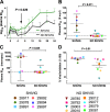
SHIVIG treatment reduces acute and set-point plasma viral load (PVL) in SHIVSF162P3-infected infant rhesus macaques. PVL for individual treated animals are compared over the period of the study for SHIVIG (n = 6) (A) and No-SHIVIG (n = 8) (B). Comparisons of PVL between SHIVIG and No-SHIVIG animals are shown at 2 weeks after challenge (C), and the area under the curve (AUC) for the 24-week study period is shown (D). Individual animals are indicated with unique symbols. P values are indicated (Mann-Whitney U test).
Fig 4
Fig 4
Effect of passively transferred SHIVIG on cell-associated virus load (CAVL). Copies of viral DNA per μg of PBMC cellular DNA were quantified by real-time PCR, and AUC values for weeks 0 to 24 were quantified for individual macaques. (A) Correlation between CAVL and PVL (vRNA per ml of plasma), P value, and Spearman rank coefficient (r) value are noted. (B) Mean CAVL in SHIVIG versus No-SHIVIG macaques during the 24 weeks after initiation of infection (w.p.i.). The horizontal dotted line shows the limit of detection of the assay. The P value is indicated (Mann-Whitney U test).
Fig 5
Fig 5
Comparison of neutralizing antibody responses in SHIVIG and No-SHIVIG macaques. (A) Mean NAb titers for the entire 24-week period for SHIVIG and No-SHIVIG animals against SHIVSF162P3 clone MC-3, assayed by the TZM-bl neutralization assay. Only the infected and Mamu-B*08- and Mamu-B*17-negative animals were considered in these analyses (n = 14). ID50 is the reciprocal of the plasma dilution necessary to inhibit infection by 50%. The horizontal dotted line shows the limit of detection of the assay. The arrow indicates the time for the initiation of the endogenous NAb response. The P value is indicated (Mann-Whitney U test). Data shown for individual animals are ID50 values at week 6 (B), ID50 values at necropsy (C), and plasma ADCVI activity as percent viral inhibition at week 6 (D).
Fig 6
Fig 6
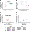
Correlations between plasma virus load and NAbs, antigen-specific (SF162 gp140) memory B cells, and B cell frequencies for macaques that developed NAbs and those that did not. (A) Plasma viral loads between weeks 8 and 24; (B) inverse correlation of PVL (weeks 8 to 24) de novo NAb development (weeks 6 to 24) during set-point viremia. Correlations of HIV-1SF162 gp140-specific memory B cells were determined by ELISPOT assay with plasma viral loads measured at the time of necropsy (C) and with total peripheral CD20+ B cell frequencies, quantified by flow cytometry, at necropsy (D). NAb+ and NAb animals are identified individually. Analysis includes only infected Mamu-B*08- and Mamu-B*17-negative animals (n = 14). Spearman correlation was used, and r and P values are given.
Fig 7
Fig 7
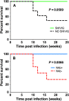
Kaplan-Meier survival curves for SHIVSF162P3-infected infant macaques. (A) Four NAb/No-SHIVIG animals were euthanized by week 16 for humane reasons due to severe disease progression (Table 1). Analysis includes only infected Mamu-B*008:01- and Mamu-B*017:01-negative animals (n = 14). (B) NAb+ and NAb animals are identified in Table 1. Log-rank (Mantel-Cox) test was used to compare survival curves. P values are indicated.
Fig 8
Fig 8
MHC-I alleles promote endogenous NAb responses. Comparison during acute infection (weeks 0 to 8) of No-SHIVIG animals (Mamu-B*08 and Mamu-B*17 positive and negative) and SHIVIG animals (Mamu-B*08 and Mamu-B*17 negative) for PVL (A) and peripheral CD20+ B cells (B). (C) Inverse correlation of acute viremia and endogenous de novo NAb response (weeks 6 to 24) and (D) comparison of endogenous NAb responses between the three groups. The P values and the correlation coefficient (r) are stated. Bonferroni and Tukey-Kramer adjustment for multiple-error correction was used to determine the significance of multiple-group (3 or more) comparisons.

References

    1. Gray ES, Madiga MC, Hermanus T, Moore PL, Wibmer CK, Tumba NL, Werner L, Mlisana K, Sibeko S, Williamson C, Abdool Karim SS, Morris L. 2011. The neutralization breadth of HIV-1 develops incrementally over four years and is associated with CD4+ T cell decline and high viral load during acute infection. J. Virol. 85:4828–4840 - PMC - PubMed
    1. Richman DD, Wrin T, Little SJ, Petropoulos CJ. 2003. Rapid evolution of the neutralizing antibody response to HIV type 1 infection. Proc. Natl. Acad. Sci. U. S. A. 100:4144–4149 - PMC - PubMed
    1. Sather DN, Armann J, Ching LK, Mavrantoni A, Sellhorn G, Caldwell Z, Yu X, Wood B, Self S, Kalams S, Stamatatos L. 2009. Factors associated with the development of cross-reactive neutralizing antibodies during human immunodeficiency virus type 1 infection. J. Virol. 83:757–769 - PMC - PubMed
    1. Wei X, Decker JM, Wang S, Hui H, Kappes JC, Wu X, Salazar-Gonzalez JF, Salazar MG, Kilby JM, Saag MS, Komarova NL, Nowak MA, Hahn BH, Kwong PD, Shaw GM. 2003. Antibody neutralization and escape by HIV-1. Nature 422:307–312 - PubMed
    1. Burton DR, Pyati J, Koduri R, Sharp SJ, Thornton GB, Parren PWHI, Sawyer LSW, Hendry RM, Dunlop N, Nara PL, Lamacchia M, Garratty E, Stiehm ER, Bryson YJ, Cao Y, Moore JP, Ho DD, Barbas CF., III 1994. Efficient neutralization of primary isolates of HIV-1 by a recombinant human monoclonal antibody. Science 266:1024–1027 - PubMed

Publication types

MeSH terms