. 2013:8:2453-63.
doi: 10.2147/IJN.S46357. Epub 2013 Jul 11.

Novel thermosensitive hydrogel for preventing formation of abdominal adhesions

Affiliations

Novel thermosensitive hydrogel for preventing formation of abdominal adhesions

Xiang Gao et al. Int J Nanomedicine. 2013.

Abstract

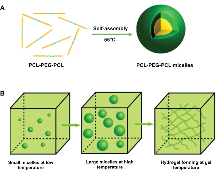
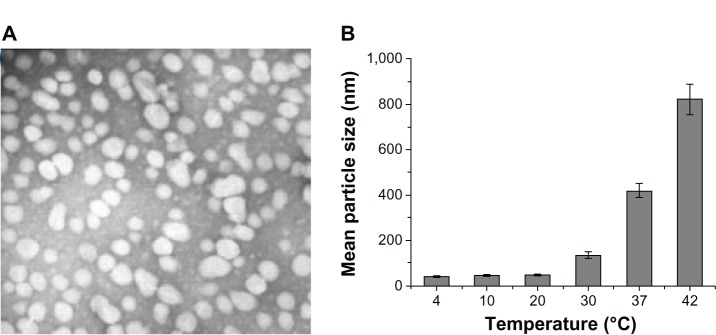
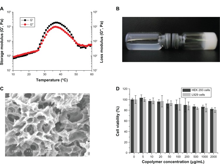
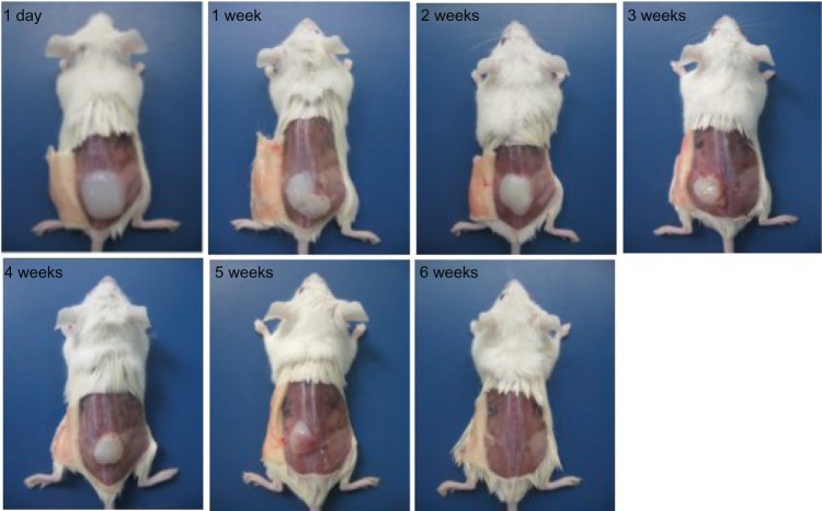
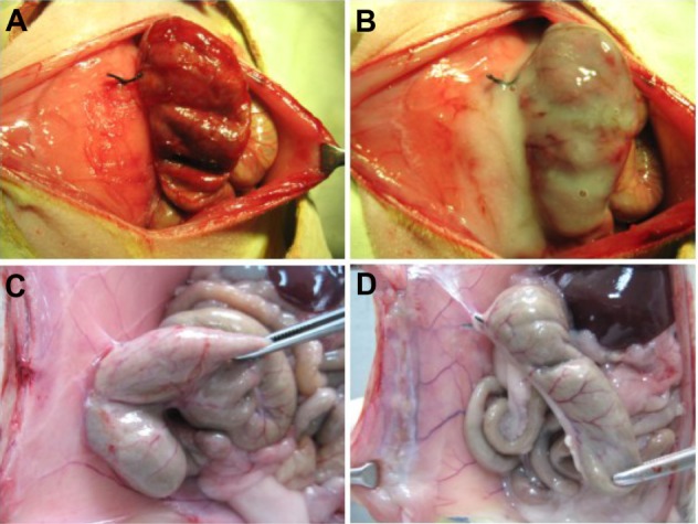
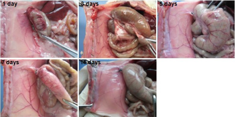
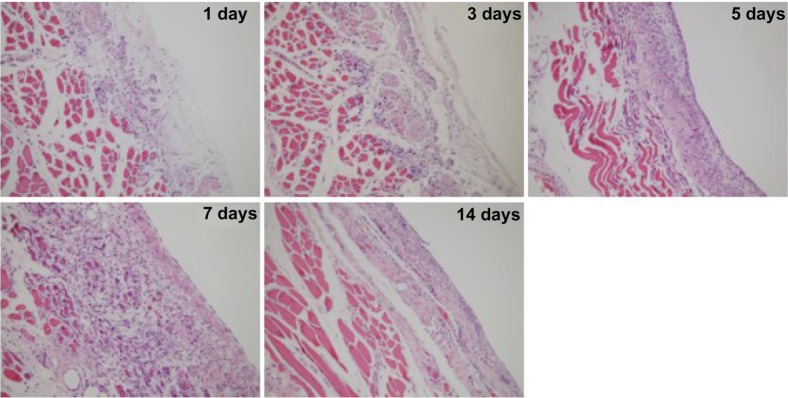
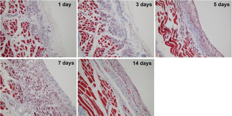
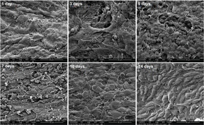
Adhesions can form after almost any type of abdominal surgery. Postoperative adhesions can be prevented by improved surgical techniques, such as reducing surgical trauma, preventing ischemia, and avoiding exposure of the peritoneal cavity to foreign materials. Although improved surgical techniques can potentially reduce formation of adhesions, they cannot be eliminated completely. Therefore, finding more effective methods to prevent postoperative adhesions is imperative. Recently, we found that a novel thermosensitive hydrogel, ie, poly(ε-caprolactone)-poly(ethylene glycol)-poly(ε-caprolactone) (PCEC) had the potential to prevent postoperative adhesions. Using the ring-opening polymerization method we prepared a PCEC copolymer which could be dissolved and assembled at 55°C into PCEC micelles with mean size of 25 nm. At body temperature, a solution containing PCEC micelles could convert into a hydrogel. The PCEC copolymer was biodegradable and had low toxicity in vitro and in vivo. We found that most animals in a hydrogel-treated group (n = 10) did not develop adhesions. In contrast, 10 untreated animals developed adhesions that could only be separated by sharp dissection (P < 0.001). The hydrogel could adhere to peritoneal wounds and degraded gradually over 7-9 days, transforming into a viscous fuid that was completely absorbed within 12 days. The injured parietal and visceral peritoneum remesothelialized over about seven and nine days, respectively. This study confirms that PCEC hydrogel has potential application in the prevention of postoperative adhesions.

Keywords: biodegradable; hydrogel; poly(ε-caprolactone)-poly(ethylene glycol)-poly(ε-caprolactone); postoperative adhesions; thermosensitive.


Figures

Figure 1
Figure 1
Preparation of thermosensitive PCEC hydrogel. (A) First, PCEC triblock copolymers were assembled into PCEC micelles at 55°C. These micelles were then stored at 4°C. (B) When PCEC micelles were used, the micelles became larger with increasing temperature, and the hydrogel formed when the temperature reached gelation temperature. Abbreviation: PCEC, poly(ε-caprolactone)(PCL)-poly(ethylene glycol)(PEG)-poly(ε-caprolactone).
Figure 2
Figure 2
(A) Transmission electron micrograph of PCEC micelles. (B) Relationship between size of micelles and temperature, with the mean size of the PCEC micelles becoming larger with increasing temperature. Abbreviation: PCEC, poly(ε-caprolactone)-poly(ethylene glycol)-poly(ε-caprolactone).
Figure 3
Figure 3
Characterization of PCEC hydrogel. (A) Rheologic analysis of PCEC-micelles-PCEC-hydrogel dual-drug delivery system as a function of temperature. (B) Morphology of blank hydrogel (20 wt%) at 4°C and 37°C. (C) Scanning electron micrograph of PCEC hydrogel. (D) Cytotoxicity of the PCEC was determined by MTT assay. Abbreviations: PCEC, poly(ε-caprolactone)-poly(ethylene glycol)-poly(ε-caprolactone); MTT, 3-(4,5-Dimethylthiazol-2-yl)-2,5-diphenyltetrazolium bromide; HEK, human embryonic kidney.
Figure 4
Figure 4
Degradation of PCEC hydrogel in vivo, with the hydrogel becoming smaller with time and completely disappearing by week 6, indicating that PCEC is biodegradable. Abbreviation: PCEC, poly(ε-caprolactone)-poly(ethylene glycol)-poly(ε-caprolactone).
Figure 5
Figure 5
Creation and treatment in the rat abdominal surgery adhesion model. (A) Rat model of abdominal wall defect-cecum. (B) PCEC hydrogel applied to the injured abdominal wall and cecum. (C) Score 4 adhesion between the injured abdominal wall and cecum in a control rat, with adhesion of an intestine segment to the injured abdominal wall and adhesion of the omentum to the sutured midline incision. (D) No adhesions were observed in the group treated with the PCEC gel. Abbreviation: PCEC, poly(ε-caprolactone)-poly(ethylene glycol)-poly(ε-caprolactone).
Figure 6
Figure 6
Prevention of abdominal adhesions in a rat abrasion model. (A and B) No adhesions were formed between the abdominal wall and cecum after two weeks of treatment with poly(ε-caprolactone)-poly(ethylene glycol)-poly(ε-caprolactone) (PCEC). (C) Adhesions between the defective cecum and surrounding tissue after two weeks in the untreated group.
Figure 7
Figure 7
Photographs taken after using dry gauze to absorb viscous liquid in the abdominal cavity in hydrogel-treated rats in the days following treatment. The poly(ε-caprolactone)-poly(ethylene glycol)-poly(ε-caprolactone) (PCEC) hydrogel transformed into a solid-like membrane adhering to the injured surfaces of the abdominal wall and wrapping around the injured cecum. The injured surfaces of the cecum could still be observed after partially stripping the hydrogel membrane. With the passage of time, the hydrogel covering on the injured surfaces of the abdominal wall and cecum gradually disappeared and transformed into a viscous liquid, which was completely absorbed by 14 days, with gradual healing of the peritoneal wounds.
Figure 8
Figure 8
Histologic observations on hematoxylin and eosin staining.
Figure 9
Figure 9
Histologic observations on Masson trichrome staining.
Figure 10
Figure 10
Scanning electron micrographs of the surface of the stripped peritoneum treated with hydrogel on the days indicated. Spherical inflammatory cells appeared on the surface of the peritoneum on day 3 that increased in number during the following two days and reached a maximum on day 5. On day 7, the spherical cells became elongated and flattened, and increased in number during the following three days, reaching a maximum on day 7. At the same time, the hydrogel covering on the surface of the peritoneum reduced with time. On day 7, the hydrogel was degraded and had completely disappeared by day 10.

References

    1. Trew G. Postoperative adhesions and their prevention. Rev Gynaecol Pract. 2006;6:47–56.
    1. Menzies D, Ellis H. Intestinal obstruction from adhesions – how big is the problem? Ann R Coll Surg Engl. 1990;72:60–63. - PMC - PubMed
    1. Szabó G, Mikó I, Nagy P, et al. Adhesion formation with open versus laparoscopic cholecystectomy: an immunologic and histologic study. Surg Endosc. 2007;21:253–257. - PubMed
    1. Menzies D. Postoperative adhesions: their treatment and relevance in clinical practice. Ann R Coll Surg Engl. 1993;75:147–153. - PMC - PubMed
    1. Schnüriger B, Barmparas G, Branco BC, Lustenberger T, Inaba K, Demetriades D. Prevention of postoperative peritoneal adhesions: a review of the literature. Am J Surg. 2011;201:111–121. - PubMed

Publication types

MeSH terms

Substances