Review
. 2014 Jan:307:86-97.
doi: 10.1016/j.heares.2013.07.008. Epub 2013 Jul 22.

Psychophysics and neuronal bases of sound localization in humans

Affiliations
Review

Psychophysics and neuronal bases of sound localization in humans

Jyrki Ahveninen et al. Hear Res. 2014 Jan.

Abstract

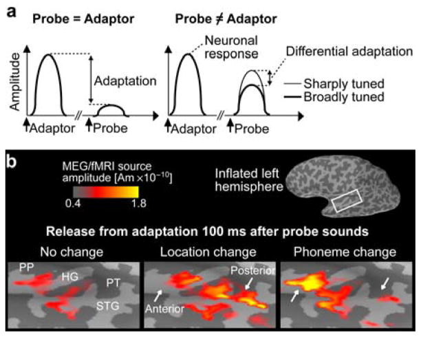
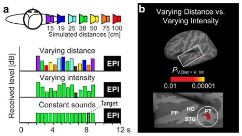
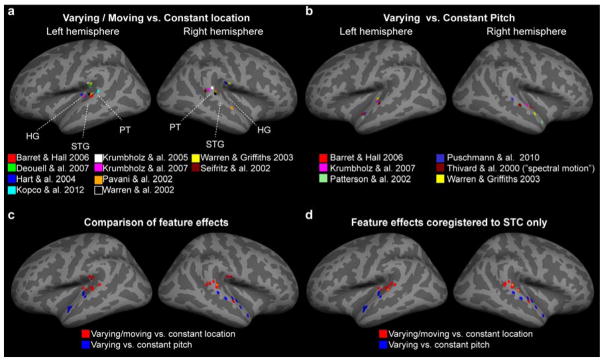
Localization of sound sources is a considerable computational challenge for the human brain. Whereas the visual system can process basic spatial information in parallel, the auditory system lacks a straightforward correspondence between external spatial locations and sensory receptive fields. Consequently, the question how different acoustic features supporting spatial hearing are represented in the central nervous system is still open. Functional neuroimaging studies in humans have provided evidence for a posterior auditory "where" pathway that encompasses non-primary auditory cortex areas, including the planum temporale (PT) and posterior superior temporal gyrus (STG), which are strongly activated by horizontal sound direction changes, distance changes, and movement. However, these areas are also activated by a wide variety of other stimulus features, posing a challenge for the interpretation that the underlying areas are purely spatial. This review discusses behavioral and neuroimaging studies on sound localization, and some of the competing models of representation of auditory space in humans. This article is part of a Special Issue entitled Human Auditory Neuroimaging.

Keywords: 3D; AC; AM; BAEP; BRIR; DRR; DTI; EEG; ERP; FM; HG; HRTF; Heschl's gyrus; ILD; IPD; ITD; MAEP; MEG; MRI; PET; PP; PT; SSR; STG; amplitude modulation; auditory cortex; binaural room impulse response; brainstem auditory evoked potentials; diffusion-tensor imaging; direct-to-reverberant ratio; electroencephalography; event-related potential; fMRI; frequency modulation; functional magnetic resonance imaging; head-related transfer function; interaural level difference; interaural phase difference; interaural time difference; magnetic resonance imaging; magnetoencephalography; middle-latency auditory evoked potential; planum polare; planum temporale; positron emission tomography; steady-state response; superior temporal gyrus; three dimensional.


Figures

Figure 1
Figure 1
Adaptation studies of auditory spatial processing. (a) Neuronal adaptation refers to suppression of responses to Probe sounds, as a function of their similarity and temporal proximity to preceding Adaptor sounds. Differential release from adaptation when Probe vs. Adaptor differences may reveal populations tuned to the varied feature dimension. (b) Our previous MEG/fMRI adaptation data (Ahveninen et al., 2006), revealing differential release from adaptation due to changes in directional vs. phonetic differences between Probe and Adaptor sounds, which supports the existence of anterior “what” and posterior “where” AC pathways (i.e., there is specific release from adaptation in posterior AC following sound location change from adaptor to probe). An fMRI-weighted MEG source estimate is shown in a representative subject.
Figure 2
Figure 2
fMRI studies on intensity-independent auditory distance processing. (a) Our recent fMRI adaptation paradigm (Kopco et al., 2012), comparing responses to sounds that are “Constant”, or contain “Varying distance” cues (all possible 3D distance cues 15–100 cm from the listener) or “Varying intensity” cues (other cues corresponding constantly to 38 cm). This kind of adaptation fMRI design presumably differentiates the tuning properties of neurons within each voxel (Grill-Spector et al., 2001), analogously to the MEG/EEG adaptation example above. (Subjects paid attention to unrelated, randomly presented duration decrements, to control attention effects.) (b) Adaptation fMRI data (Kopco et al., 2012), demonstrating a comparison between the varying distance and varying intensity conditions (N=11). Maximal difference is observed in the posterior STG/PT, possibly reflecting neurons sensitive to intensity-independent distance cues.
Figure 3
Figure 3
Adaptation fMRI meta-analysis of release from adaptation due to spatial or pitch/spectral variation. From each study, the reported voxels showing largest signals during varying location (a; motion vs. stationary, varying location vs. stationary) or pitch (b; varying vs. constant pitch) have been coregistered to the nearest cortical vertex in the Freesurfer standard brain representation. In case separate subregions, such as when PT vs. HG voxels were reported separately (Hart et al., 2004; Warren et al., 2003), the AC voxel showing the largest signal was selected. (c) The comparisons of feature effects suggest quite a clear division between the putative posterior “where” pathway (observations labeled red; orange in the case of two overlapping voxels) and the anterior areas demonstrating release from adaptation due to pitch-related variation (blue; possibly with further subregions). (d) Finally, an additional analysis where the peak voxels have been coregistered to the closest superior temporal cortex (STC) vertex is presented, to account for possible misalignment along the vertical axis across the studies. Note that Thivard and colleagues (2000) did not investigate pitch, per se. The results are shown due to their importance on influential alternative hypotheses related to “spectral motion” and PT (Belin et al., 2000; Griffiths et al., 2002). Altogether, this surface-based “adaptation-fMRI meta analysis” demonstrates the importance of consistent anatomical frameworks in evaluating the distinct AC subareas.

References

    1. Adler FH. Physiology Of The Eye. C. V. Mosby; St. Louis, MO: 1959.
    1. Adriani M, Maeder P, Meuli R, Thiran AB, Frischknecht R, Villemure JG, Mayer J, Annoni JM, Bogousslavsky J, Fornari E, Thiran JP, Clarke S. Sound recognition and localization in man: specialized cortical networks and effects of acute circumscribed lesions. Exp Brain Res. 2003;153:591–604. - PubMed
    1. Ahveninen J, Huang S, Belliveau JW, Chang WT, Hämäläinen M. Dynamic oscillatory processes governing cued orienting and allocation of auditory attention. J Cogn Neurosci. 2013 In press. - PMC - PubMed
    1. Ahveninen J, Jääskeläinen IP, Belliveau JW, Hämäläinen M, Lin FH, Raij T. Dissociable Influences of Auditory Object vs. Spatial Attention on Visual System Oscillatory Activity. PLoS One. 2012;7:e38511. - PMC - PubMed
    1. Ahveninen J, Hämäläinen M, Jääskeläinen IP, Ahlfors SP, Huang S, Lin FH, Raij T, Sams M, Vasios CE, Belliveau JW. Attention-driven auditory cortex short-term plasticity helps segregate relevant sounds from noise. Proc Natl Acad Sci U S A. 2011;108:4182–7. - PMC - PubMed

Publication types