. 2013 Sep 26;122(13):2195-204.
doi: 10.1182/blood-2013-04-496950. Epub 2013 Jul 25.

HIV-1 infection of hematopoietic progenitor cells in vivo in humanized mice

Affiliations

HIV-1 infection of hematopoietic progenitor cells in vivo in humanized mice

Christopher C Nixon et al. Blood. .

Abstract

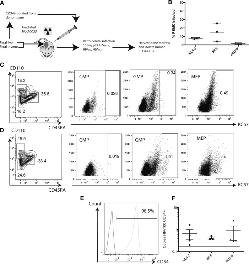
HIV infection has been associated with defective hematopoiesis since the earliest days of the HIV/AIDS epidemic. Generation of all hematopoietic lineages suffers in the face of infection. The mechanisms by which HIV impairs normal blood cell development remain unclear, and direct infection of intermediate hematopoietic progenitors has not been established as a source of HIV-associated hematopoietic pathology. Here, we demonstrate infection of multiple subsets of highly purified intermediate hematopoietic progenitors by wild-type HIV both in vitro and in vivo. Although direct infection is clearly cytotoxic, we find that some infected progenitors can survive and harbor proviral DNA. We report intermediate hematopoietic progenitors to be a novel target of infection and their permissivity to infection increases with development. Further, the nonobese diabetic severe combined immunodeficiency common γ chain knockout-bone marrow-liver-thymus humanized mouse provides a unique model for studying the impact of HIV infection on bone marrow-based human hematopoiesis.


Figures

Figure 1
Figure 1
VSV-G pseudotyped infection of CD34+ HPCs results in developmental pathology in multiple hematopoietic lineages. (A) The NL4HSA-eGFP vector is based on the NL4-3 strain of HIV-1. The vector is deficient in env and vpr because of the insertion of marker genes and was pseudotyped with VSV-G to produce viral vector particles. (B) FACS strategy for differentiating GFP-positive and GFP-negative CD34+ hematopoietic progenitor cells 48 hours after transduction. (C) Quantitative PCR analysis for copies of HIV per GFP+ cell indicates a single integrant on average for each donor. (D) Colonies grown in methylcellulose derived from NL4HSA-eGFP–transduced cells (right) are in general much smaller than those derived from nontransduced cells (left). (E) CD34+ hematopoietic progenitors transduced with NL4-HSA-eGFP generate far fewer colonies than do nontransduced cells. Each mark represents an individual methylcellulose plate and the data represent the mean ± the standard error of the mean (SEM). (F) There is a skewing of lineages in NL4-HSA-eGFP–transduced cultures compared with nontransduced. Erythroid colonies are particularly impacted. (G) Megakaryocyte colonies are likewise diminished from transduced progenitors. Each mark represents an individual plate and the data represent the mean ± SEM. LTR, long terminal repeat; SSC, side scatter.
Figure 2
Figure 2
CD34+CD38+ intermediate hematopoietic progenitors express cell surface receptors necessary for HIV-1 entry. (A) Within the CD34+ HPC population, CD38 coexpression indicates a more mature phenotype and includes the CMP, MEP, and GMP. (B) CD34+ cells were enriched from fetal liver and assayed by flow cytometry for the presence of HIV entry receptors in conjunction with definitive surface markers of intermediate hematopoietic progenitors. CD34+CD38+ double-positive cells were gated (left) to select the CMP, GMP, and MEP based on CD123 and CD45RA expression (right). (C) Each population of intermediate hematopoietic progenitor was examined for CD4 and CCR5 expression by flow cytometry. (D) These same populations were also examined for CD4 and CXCR4 expression by flow cytometry. LT-HSC, long-term hematopoietic stem cell; MPP, multipotent progenitor; ST-HSC, short-term hematopoietic stem cell.
Figure 3
Figure 3
Intermediate hematopoietic progenitors are increasingly permissive to HIV-1 infection in vitro with progressive development. (A) CD34+ cells were sorted to high purity by MACS from mononuclear cells purified from human fetal liver. Flow cytometry shows that of the live sorted cells, 98% express CD34 (left). From these, CD38+ cells were sorted to isolate each subpopulation of intermediate hematopoietic progenitors (center). The CMP, MEP, and GMP are all CD34+CD38+; the CMP expresses neither CD45RA nor CD110. Only the MEP expresses CD110; the GMP is CD45RA+CD110− (left). (B) Purity of the sorted CMP is indicated by CD110 expression (left) and CD45RA expression (center). Cells were infected with HIV89.6 and assayed by qRT-PCR 48 hours later for full-length viral DNA (right) in the presence or absence of AZT. Each mark indicates a single donor; the data represent the mean ± SEM. (C) Purity of sorted GMP indicated by CD110 expression (left) and CD45RA expression (center). (D) Purity of the sorted MEP indicated by CD110 expression (left) and CD45RA expression (center). FSC, forward scatter; SSC, side scatter.
Figure 4
Figure 4
Intermediate HPCs derived from the bone marrow of HIV-infected NSG-BLT mice are productively infected as shown by HIV-GAG expression. (A) Schematic of experimental design for infecting humanized NSG-BLT mice and isolating CD34+ HPCs from their bone marrow. (B) Peripheral blood mononuclear cells from infected mice were analyzed by PCR for the presence of full-length viral DNA relative to the number of copies of human β-globin to determine the percent of infected cells. Each mark represents the average of 3 reactions for each mouse; the total data represent the pooled mean ± SEM of all reactions from all mice. Cells isolated from the murine bone marrow were analyzed for the presence of human intermediate HPC markers in conjunction with HIV-GAG expression. (C) Cells derived from uninfected mice. (D) Cells derived from mice exposed to HIVNL4-3. (E) Representative flow cytometric analysis of CD34+ purity after sorting from BLT bone marrow. (Left) Isotype control; (right) CD34 staining at 98.5% purity. (F) Human CD34+ HSC derived from BLT bone marrow harbor full-length HIV DNA when analyzed by quantitative PCR. Each mark indicates the average of triplicate assays in a single mouse; the total data represent the pooled mean ± SEM of all reactions from all mice. The dashed line indicates the limit of detection of the assay. JRCSF, the patient code used to define the particular strain of HIV.
Figure 5
Figure 5
HPCs from HIV-infected NSG-BLT mice generate colonies that harbor proviral DNA. (A) The total number of colonies that develops from each infected mouse is significantly less than those from uninfected mice. Each mark represents the average of triplicate assays in a single mouse; the total data represent the pooled mean ± SEM from all mice. (B) Phenotypic analysis of colonies indicates significant differences in lineage commitment from HSC derived from infected mice compared with uninfected mice. (C) Erythroid lineage development is particularly impaired in the HSC derived from HIV-1–infected mice. Each mark represents the average of triplicate assays in a single mouse; the total data represent the pooled mean ± SEM from all mice. (D) Colonies derived from human CD34+ cells isolated from the bone marrow of infected and uninfected mice were assayed by qRT-PCR for the presence of full-length viral DNA. Colonies were first phenotyped visually by light microscopy. E, erythroid; G, granulocyte; GM, granulocyte-macrophage mixed; M, macrophage. Percentages are of that particular colony type in which HIV was detected compared with the total number of that colony type assayed. (E) HIV+ colonies were assayed by alu-Gag PCR to determine integration site. Following successful amplification, amplicons were sequenced to determine the gene into which the virus integrated. Four different mice were assayed, representing infection by each of the 3 viral strains.

Comment in

References

    1. Rosenfeld SJ, Young NS. Viruses and bone marrow failure. Blood Rev. 1991;5(2):71–77. - PubMed
    1. Marche C, Tabbara W, Michon C, Clair B, Bricaire F, Matthiessen L. Bone marrow findings in HIV infection: a pathological study. Prog AIDS Pathol. 1990;2:51–60. - PubMed
    1. Donahue RE, Johnson MM, Zon LI, Clark SC, Groopman JE. Suppression of in vitro haematopoiesis following human immunodeficiency virus infection. Nature. 1987;326(6109):200–203. - PubMed
    1. Treacy M, Lai L, Costello C, Clark A. Peripheral blood and bone marrow abnormalities in patients with HIV related disease. Br J Haematol. 1987;65(3):289–294. - PubMed
    1. Scaradavou A. HIV-related thrombocytopenia. Blood Rev. 2002;16(1):73–76. - PubMed

Publication types

LinkOut - more resources