. 2013 Nov:90:40-7.
doi: 10.1016/j.mvr.2013.06.012. Epub 2013 Jul 22.

Angiogenic endothelial cell invasion into fibrin is stimulated by proliferating smooth muscle cells

Affiliations

Angiogenic endothelial cell invasion into fibrin is stimulated by proliferating smooth muscle cells

Areck A Ucuzian et al. Microvasc Res. 2013 Nov.

Abstract

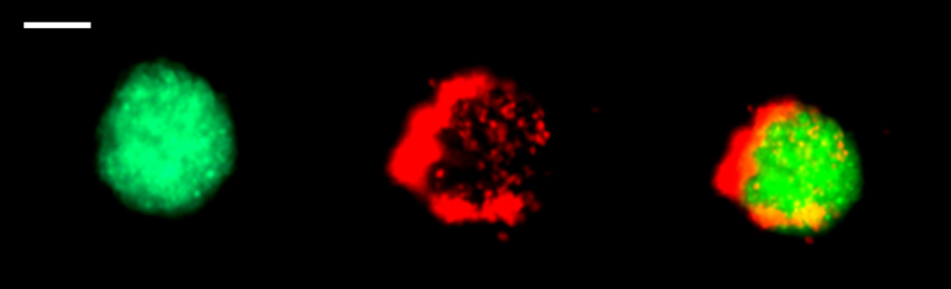
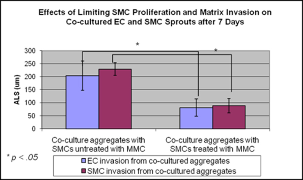
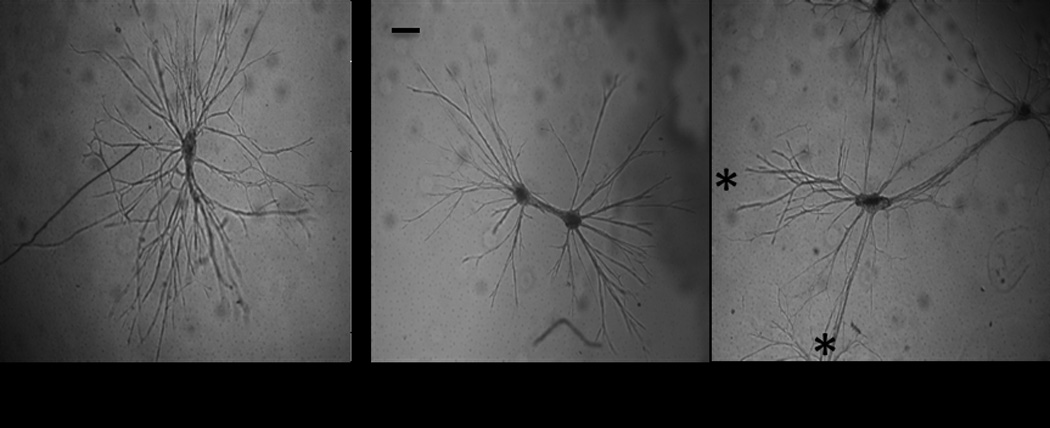
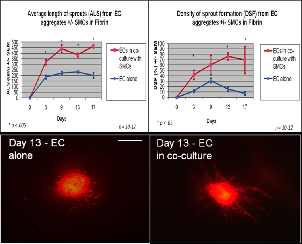
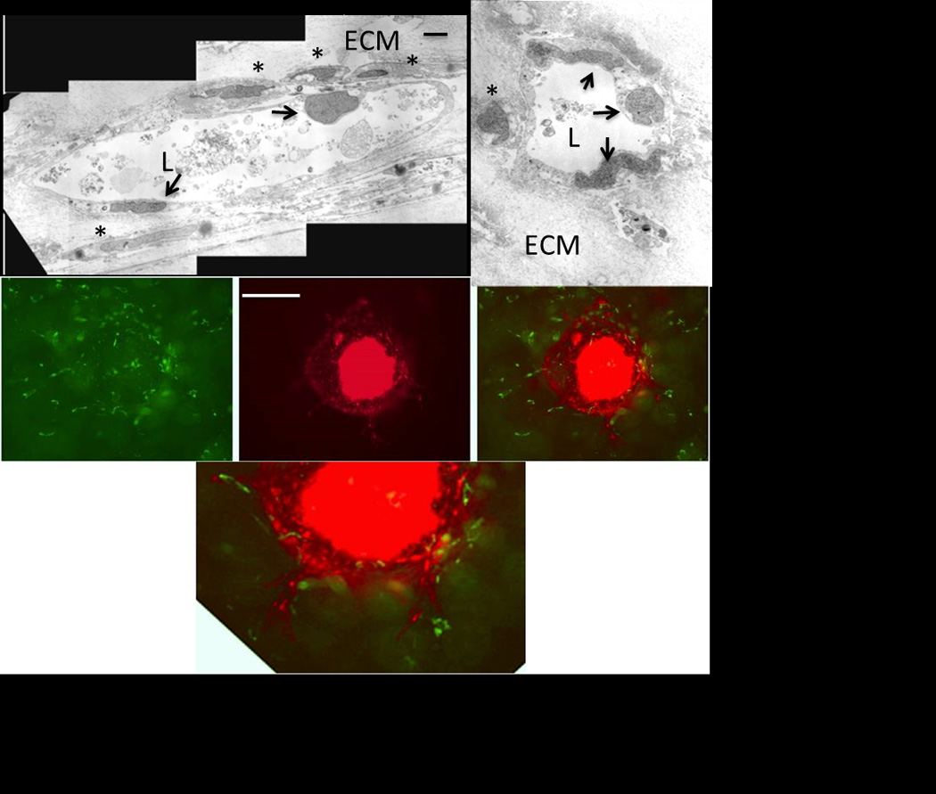
These studies aimed to determine the effect of smooth muscle cells (SMCs) on angiogenic behavior of endothelial cells (ECs) within fibrin hydrogels, an extracellular matrix (ECM) commonly used in tissue engineering. We developed a 3-D, fibrin-based co-culture assay of angiogenesis consisting of aggregates of SMCs with ECs seeded onto the aggregates' surface. Using digital fluorescence micrography, EC matrix invasion was quantified by average length of sprouts (ALS) and density of sprout formation (DSF). We demonstrated that ECs and SMCs co-invade into the ECM in close proximity to one another. ECs that were co-cultured with SMCs demonstrated increased invasion compared to ECs that were cultured alone at all time points. At Day 19, the ALS of ECs in co-culture was 327+/-58μm versus 70+/-11μm of ECs cultured alone (p=.01). The DSF of co-cultured ECs was also significantly greater than that of ECs cultured alone (p=.007 on Day 19). This appeared to be a function of both increased EC invasion as well as improved persistence of EC sprout networks. At 7days, ECs in co-culture with proliferation-inhibited SMCs previously treated with Mitomycin-C (MMC) demonstrated significantly attenuated sprouting compared to ECs co-cultured with SMCs that were untreated with MMC (82+/-14μm versus 205+/-32μm; p<.05). In assays in which multiple co-culture aggregates were cultured within a single hydrogel, we observed directional invasion of sprouts preferentially towards the other aggregates within the hydrogel. In co-culture assays without early EC/SMC contact, the ALS of ECs cultured in the presence of SMCs was significantly greater than those cultured in the absence of SMCs by Day 3 (320+/-21μm versus 187+/-16μm; p<.005). We conclude that SMCs augment EC matrix invasion into 3-D fibrin hydrogels, at least in part resulting from SMC proliferative and invasive activities. Directed invasion between co-culture aggregates and augmented angiogenesis in the absence of early contact suggests a paracrine mechanism for the observed results.


Figures

Figure 1
Figure 1
Co-culture assay #1. Left) Fluorescently green labeled SMC aggregate. Middle) Fluorescently labeled red ECs coating SMC aggregate. Right) Merged image of red ECs covering the surface of the green labeled SMC aggregate. Mag.10x. Bar 100 µm.
Figure 2
Figure 2
Quantification of angiogenesis. Mag 4x. ALS=Average length of sprouts; DSF=Density of sprout formation; L=Length; R=Radius of EC pellet.
Figure 3
Figure 3
EC sprouts in the presence and absence of SMCs. Only EC sprouts are shown for simplification.(Mag. 4x) Bar 200µm.
Figure 4
Figure 4
Quantification of EC sprouting in co-culture angiogenesis assay. ECs were cultured in the presence and absence of SMCs in direct contact with SMCs and the average length of sprouts (left) and density of sprout formation was quantified (right). N = 5; *p < 0.05.
Figure 5
Figure 5
Invasion of ECs and SMCs into fibrin from co-culture aggregates. Left) ECs (red) co-invade with SMCs (green) into fibrin hydrogels (Mag. 10x). Bar 200 µm. Right) Average length of EC and SMC sprouts from a co-culture aggregate. N = 5; *p < 0.05.
Figure 6
Figure 6
Inhibiting SMC proliferation with MMC inhibits both SMC and EC matrix invasion into fibrin hydrogels in the co-culture angiogenesis model. N = 5; *p < 0.05.
Figure 7
Figure 7
Directional invasion of co-culture sprouts. (Left) Sprouts from individual co-culture aggregates demonstrate random invasion into the hydrogel. (Middle and right panels) Sprouts from co-culture aggregates demonstrate directional invasion towards other co-culture aggregates embedded within the same hydrogels. The two right panels demonstrate two separate hydrogels, the middle panel has two aggregates embedded within the hydrogel, the right panel has five (three seen). Asterisks represent location of the two aggregates embedded within the hydrogel which were out of the field of view. Magnification, 4x. Bar 200 µm.
Figure 8
Figure 8
Top) Quantification of angiogenesis in the presence and absence of SMCs (400,000 SMCs/mL). Bottom) Angiogenesis in the presence and absence of SMCs (400,000 SMCs/mL). Only ECs shown for simplification (Mag. 4x). n = 10–12; * p < 0.05. Bar 200 µm.
Figure 9
Figure 9
Top) Cross sectional morphology of co-culture sprouts by transmission electron microscopy after 7 days of co-culture. Selected images demonstrate EC lined luminal structures with SMCs apposed to the abluminal surface of the capillary. The sprouts and associated cells were completely surrounded by the fibrin extracellular matrix. Debris was present within the lumen of the sprouts in each of the sections. Bar 1 µm. ECM = extracellular matrix; L = Lumen; Arrows = ECs; * = SMCs. Bottom) Sprouting from co-culture assay at 7 days. Left) Fluorescently labeled green SMCs are homogenously suspended within the fibrin hydrogel. Middle) Fluorescently red labeled ECs are seen sprouting from a central aggregate of ECs into the matrix. Right) Merged image of ECs sprouting into the ECM containing SMCs which associate with EC sprouts. Mag 10x. (Lower image) Digital zoom of merged image. Mag 20x. Bar 200 µm.

References

    1. Bergers G, Song S, Meyer-Morse N, Bergsland E, Hanahan D. "Benefits of targeting both pericytes and endothelial cells in the tumor vasculature with kinase inhibitors". The Journal of clinical investigation. 2003;111(9):1287. - PMC - PubMed
    1. Brewster LP, Brey EM, Tassiopoulos AK, Xue L, Maddox E, Armistead D, Burgess WH, Greisler HP. "Heparin-independent mitogenicity in an endothelial and smooth muscle cell chimeric growth factor (S130K-HBGAM)". Am J Surg. 2004;188(5):575–579. - PubMed
    1. Brewster LP, Ucuzian AA, Brey EM, Liwanag M, Samarel AM, Greisler HP. "FRNK overexpression limits the depth and frequency of vascular smooth muscle cell invasion in a three-dimensional fibrin matrix". J Cell Physiol. 225(2):562–568. - PMC - PubMed
    1. Brey EM, McIntire LV, Johnston CM, Reece GP, Patrick CW., Jr. "Three-dimensional, quantitative analysis of desmin and smooth muscle alpha actin expression during angiogenesis". Annals of Biomedical Engineering. 2004;32(8):1100. - PubMed
    1. Chon JH, Wang HS, Chaikof EL. "Role of fibronectin and sulfated proteoglycans in endothelial cell migration on a cultured smooth muscle layer". The Journal of surgical research. 1997;72(1):53. - PubMed

LinkOut - more resources