. 2013 Aug;8(8):1583-601.
doi: 10.1038/nprot.2013.094. Epub 2013 Jul 25.

Design and generation of recombinant rabies virus vectors

Affiliations

Design and generation of recombinant rabies virus vectors

Fumitaka Osakada et al. Nat Protoc. 2013 Aug.

Abstract

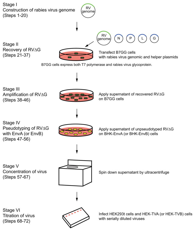
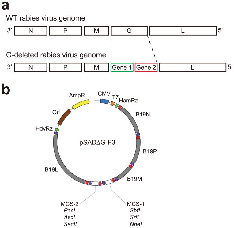
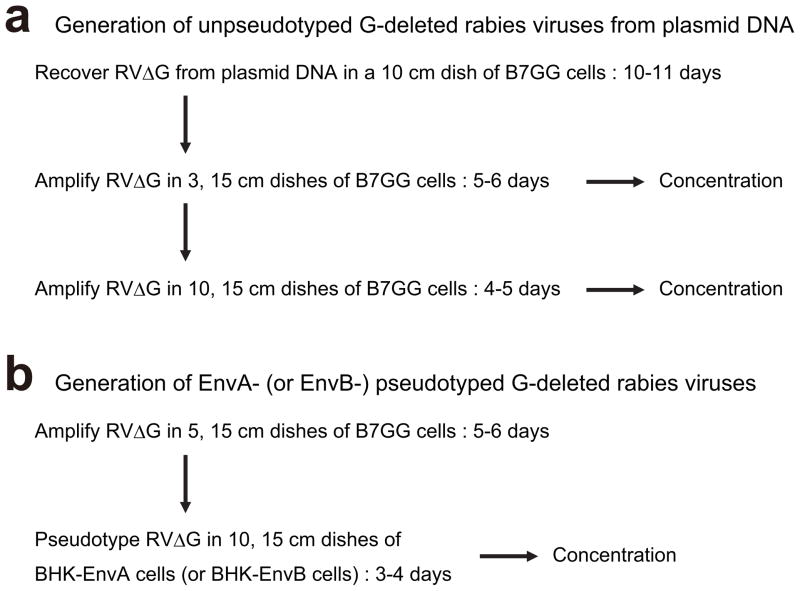
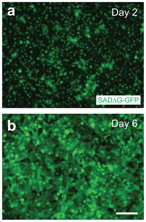
Rabies viruses, negative-strand RNA viruses, infect neurons through axon terminals and spread trans-synaptically in a retrograde direction between neurons. Rabies viruses whose glycoprotein (G) gene is deleted from the genome cannot spread across synapses. Complementation of G in trans, however, enables trans-synaptic spreading of G-deleted rabies viruses to directly connected, presynaptic neurons. Recombinant rabies viruses can encode genes of interest for labeling cells, controlling gene expression and monitoring or manipulating neural activity. Cre-dependent or bridge protein-mediated transduction and single-cell electroporation via the EnvA-TVA or EnvB-TVB (envelope glycoprotein and its specific receptor for avian sarcoma leukosis virus subgroup A or B) system allow cell type-specific or single cell-specific targeting. These rabies virus-based approaches permit the linking of connectivity to cell morphology and circuit function for particular cell types or single cells. Here we describe methods for construction of rabies viral vectors, recovery of G-deleted rabies viruses from cDNA, amplification of the viruses, pseudotyping them with EnvA or EnvB and concentration and titration of the viruses. The entire protocol takes 6-8 weeks.


Figures

Figure 1
Figure 1. Flowchart and timeline for G-deleted rabies virus production
Generation of G-deleted rabies virus (RVΔG) consist of 6 stages: (a) construction of rabies viral genome, (b) recovery of G-deleted rabies virus from plasmids, (c) amplification of G-deleted rabies virus, (d) pseudotyping with EnvA or EnvB, (e) concentration of virus, and (f) titration of virus.
Figure 2
Figure 2. Rabies virus genome and G-deleted rabies virus vector
(a) Genomes of wild type rabies virus and G-deleted rabies virus. N: nucleoprotein. P: phosphoprotein. M: matrix protein. G: glycoprotein. L: large protein. (b) Map of the G-deleted rabies virus genomic vector pSADΔG-F3. All unique cloning sites are indicated. Red and purple boxes indicate transcription start and stop sequences, respectively. Further information on pSADΔG-F3 is provided in a previous publication by Osakada et al..
Figure 3
Figure 3. Typical scheme for G-deleted rabies virus production
(a) Generation of G-deleted rabies virus from plasmid DNA. Recovery of G-deleted rabies virus from plasmids takes 10–12 days. Virus can be concentrated and frozen at each step. Recovered virus is typically amplified two times for concentration to get enough aliquots of the same batch. Recovery, amplification and concentration of virus correspond to steps 21–36, 37–46, and 57–67, in the text, respectively. (b) Pseudotyping of G-deleted rabies virus with EnvA or EnvB. Pseudotyping requires preparing unpseudotyped G-deleted rabies virus in advance. Pseudotyping and concentration correspond to steps 47–56 and 57–67 in the text, respectively.
Figure 4
Figure 4. Recovery of G-deleted rabies viruses from DNA plasmid
Photomicrographs represent typical examples of recovery of SADΔG-GFP on day 4 (A), day 6 (B), day 8 (C) and day 10 (D). Nuclear GFP is derived from B7GG cells, whereas cytosolic GFP is derived from SADΔG-GFP. Scale bar, 300 μm.
Figure 5
Figure 5. Amplification of G-deleted rabies viruses in B7GG cells
Photomicrographs represent typical examples of amplification of SADΔG-GFP on day 2 (A), and day 6 (B). Nuclear GFP is derived from B7GG cells, whereas cytosolic GFP is derived from SADΔG-GFP. Scale bar, 100 μm.

References

    1. Luo L, Callaway EM, Svoboda K. Genetic dissection of neural circuits. Neuron. 2008;57:634–660. - PMC - PubMed
    1. Arenkiel BR, Ehlers MD. Molecular genetics and imaging technologies for circuit-based neuroanatomy. Nature. 2009;461:900–907. - PMC - PubMed
    1. Scanziani M, Hausser M. Electrophysiology in the age of light. Nature. 2009;461:930–939. - PubMed
    1. Yizhar O, Fenno LE, Davidson TJ, Mogri M, Deisseroth K. Optogenetics in neural systems. Neuron. 2011;71:9–34. - PubMed
    1. Carandini M, Heeger DJ. Normalization as a canonical neural computation. Nat Rev Neurosci. 2011;13:51–62. - PMC - PubMed

Publication types