. 2013 Sep-Oct;4(5):361-70.
doi: 10.4161/gmic.25824. Epub 2013 Jul 24.

Molecular detection of bacterial contamination in gnotobiotic rodent units

Affiliations

Molecular detection of bacterial contamination in gnotobiotic rodent units

Christopher D Packey et al. Gut Microbes. 2013 Sep-Oct.

Abstract

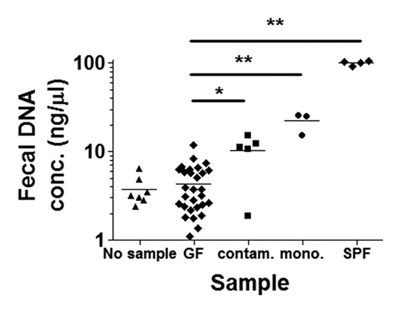
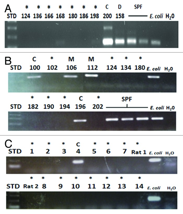
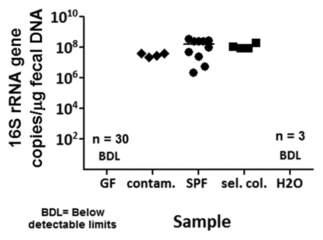
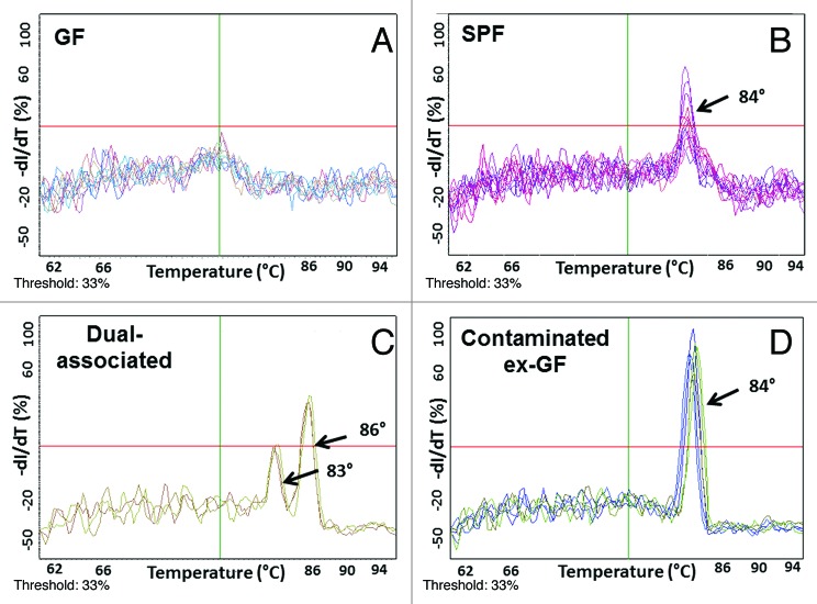
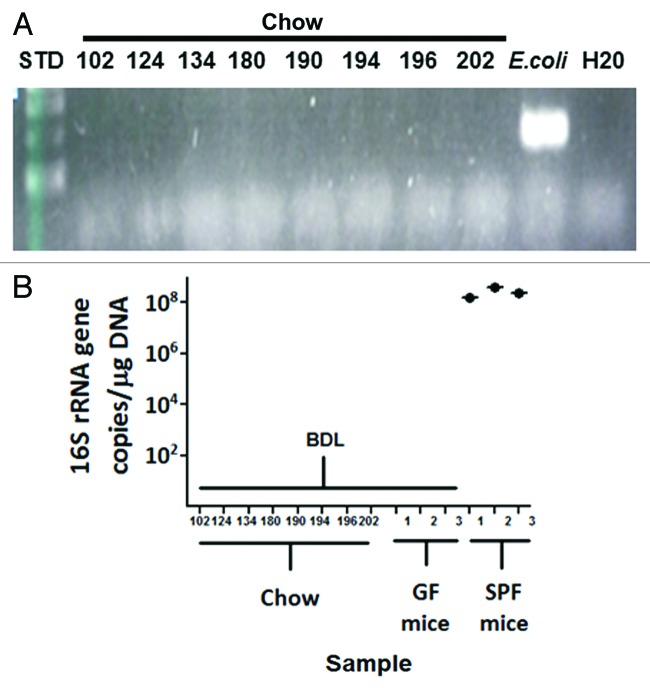
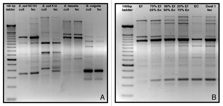
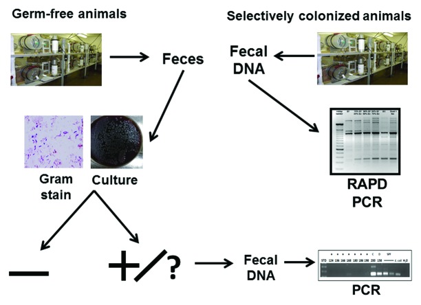
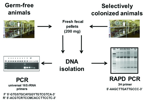
Gnotobiotic rodents provide an important technique to study the functional roles of commensal bacteria in host physiology and pathophysiology. To ensure sterility, these animals must be screened frequently for contamination. The traditional screening approaches of culturing and Gram staining feces have inherent limitations, as many bacteria are uncultivable and fecal Gram stains are difficult to interpret. Thus, we developed and validated molecular methods to definitively detect and identify contamination in germ-free (GF) and selectively colonized animals. Fresh fecal pellets were collected from rodents housed in GF isolators, spontaneously contaminated ex-GF isolators, selectively colonized isolators and specific pathogen-free (SPF) conditions. DNA isolated from mouse and rat fecal samples was amplified by polymerase chain reaction (PCR) and subjected to quantitative PCR (qPCR) using universal primers that amplify the 16S rRNA gene from all bacterial groups. PCR products were sequenced to identify contaminating bacterial species. Random amplification of polymorphic DNA (RAPD) PCR profiles verified bacterial inoculation of selectively colonized animals. These PCR techniques more accurately detected and identified GF isolator contamination than current standard approaches. These molecular techniques can be utilized to more definitively screen GF and selectively colonized animals for bacterial contamination when Gram stain and/or culture results are un-interpretable or inconsistent.

Keywords: 16S rRNA; PCR; RAPD PCR; commensal bacteria; gnotobiotic; microbiota; qPCR.


Figures

None
Figure 1. Fecal DNA concentrations as measured by spectrophotometry. The fecal DNA concentrations in putative germ-free (GF) rodents, represented by diamonds in the second lane from the left, are significantly different from fecal DNA concentrations in spontaneously contaminated ex-GF rodents (squares; middle lane). However, there was slight overlap in the respective fecal DNA concentrations between these two groups. This rendered spectrophotometry insufficient as a screening tool for bacterial GF contamination. Differences in fecal DNA concentrations between putative GF and monoassociated (circles; second from right) and specific pathogen-free (SPF) (diamonds; far right) mice were also significant. No sample, DNA extraction protocol run without sample present; contam., spontaneously contaminated, ex-GF; mono., monoassociated; *p = 0.0002; **p = 0.0001.
None
Figure 2. Agarose gel electrophoresis of PCR products of DNA isolated from rodent feces. (A) 8 National Gnotobiotic Rodent Resource Center (NGRRC) isolators that were thought to be GF were screened with 16S rRNA (rRNA) polymerase chain reaction (PCR) of mouse fecal DNA. Bacterial contamination was detected in isolator 200. Dual-associated mouse fecal DNA from isolator 158, SPF mouse fecal DNA and Escherichia coli genomic DNA served as positive controls for these PCR assays. (B) 12 NGRRC gnotobiotic isolators were blindly screened with 16S rRNA PCR of mouse fecal DNA. Fecal DNA isolated from mice from four isolators amplified: Fecal DNA from mice in isolators 106 and 112, which were subsequently revealed to be monoassociated and fecal DNA from mice in isolators 100 and 196, which were ex-GF isolators that had been recently determined to be spontaneously contaminated. SPF mouse fecal DNA and E. coli genomic DNA served as positive controls for these PCR assays. (C) 14 mouse (lanes marked 1–14) and two rat (lanes marked Rat1 and Rat2) isolators at the Gnotobiotic Animal Core (GAC) of the Center for Gastrointestinal Biology and Disease (CGIBD) at North Carolina State University were blindly screened with 16S rRNA PCR of rodent fecal DNA. The GF status of 15 of these isolators was confirmed, as was the recently-contaminated status of ex-GF isolator #4. E. coli genomic DNA served as positive controls for these PCR assays. STD, 100 base pair ladder; *, GF; C, spontaneously contaminated ex-GF; D, dual-associated; E. coli, E. coli genomic DNA; H20, water negative control template; M, monoassociated.
None
Figure 3. Bacterial levels in gnotobiotic rodent fecal samples determined by qPCR. Quantitative PCR (qPCR) assays were utilized to determine bacterial levels in gnotobiotic fecal samples by quantitating copies of the 16S rRNA gene per microgram of fecal DNA. The 16S rRNA gene was not amplified to detectable levels in GF mice (n = 30). There were on the order of 10–10 copies of the 16S rRNA gene per microgram of fecal DNA in spontaneously contaminated ex-GF mice (n = 4), SPF mice (n = 10) and selectively colonized mice (n = 4). BDL, below detectable limits; contam., contaminated ex-GF; sel. col., selectively colonized; H20, water negative control template.
None
Figure 4. 16S rRNA qPCR melting curves from gnotobiotic and SPF mouse fecal DNA. Melting curves from qPCR assays reveal that true amplification of the 16S rRNA gene occurs at melting temperatures of 83° to 87°C (B-D). No amplification from GF mouse fecal DNA is seen at these melting temperatures (A).
None
Figure 5. PCR and qPCR screening of animal chow for bacterial DNA. DNA was isolated from several types of chow used in various NGRRC isolators, and there was no detectable amplification from these samples with 16S rRNA PCR (A) or qPCR (B).E. coli genomic DNA served as a positive control for the PCR assay.
None
Figure 6. Distinguishing different individual bacterial strains by RAPD PCR fingerprints. (A) Unique rapid amplification of polymorphic DNA (RAPD) fingerprints of DNA isolated from pure cultures of E. coli NC101, E. coli K12, Enterococcus faecalis OG1RF and Bacteroides vulgatus exhibited a high degree of similarity to fingerprints of fecal DNA from rodents monoassociated with each of these bacterial strains. (B) Inoculation of E. coli NC101 monoassociated mice with E. faecalis OG1RF (and vice versa) was easily determined using RAPD PCR. The 2nd lane from the left shows RAPD PCR products from DNA isolated from pure culture of E. faecalis. The second lane from the right shows RAPD PCR products from DNA isolated from pure culture of E. coli. Lanes 3, 4 and 5 show mixtures of RAPD PCR products of E. faecalis and E. coli pure culture DNA. The 50/50 mixture of E. faecalis and E. coli DNA RAPD PCR products in lane 4 matches the fingerprint of DNA isolated from E. faecalis/E. coli dual-associated mice, shown in the far right lane. Lanes 3 and 5 show RAPD PCR profiles of DNA isolated from 75/25 mixtures of E. faecalis/E. coli and E. coli/E. faecalis, respectively. Cult, pure culture DNA; fec, fecal DNA; Ef, Enteroccous faecalis pure culture DNA; Ec, E. coli pure culture DNA.
None
Figure 7. Distinguishing different bacterial strains in complex microbial communities using RAPD PCR. The 7 bacterial strains belonging to the SIHUMI cocktail demonstrated unique fingerprints. Each of these fingerprints was clearly identifiable when up to 4 constituent strains were mixed. From left to right after the ladder on the left side, E. coli (blue), Ruminococcus gnavus (red), E. faecalis (green) and Bifidobacterium longum (orange) are shown, followed by combinations of two, three or four of the respective strains. Unique bands for each bacterium are highlighted by appropriately colored boxes, including two bands that are unique to E. coli.
None
Figure 8. Gnotobiotic unit contamination screening algorithm. The continued use of inexpensive Gram staining and culturing for frequent screening of bacterial contamination of GF animals is recommended. However, when Gram stain or culture results are difficult to interpret or inconsistent, a compromise in a glove, sleeve, or isolator wall occurs, an autoclave malfunction is diagnosed, or a large scale experiment is planned, it is recommended that these PCR and qPCR techniques be used to definitively determine the presence or absence of contamination in GF units. RAPD PCR can be used to screen selectively colonized animals for contamination.
None
Figure 9. Experimental design. DNA was isolated from GF rodent fecal samples and used as templates for PCR assays using universal bacterial 16S rRNA primers. DNA was similarly isolated from selectively colonized rodent fecal samples and used as templates for RAPD PCR assays.

References

    1. Packey CD, Sartor RB. Commensal bacteria, traditional and opportunistic pathogens, dysbiosis and bacterial killing in inflammatory bowel diseases. Curr Opin Infect Dis. 2009;22:292–301. doi: 10.1097/QCO.0b013e32832a8a5d. - DOI - PMC - PubMed
    1. Ivanov II, Frutos RdeL, Manel N, Yoshinaga K, Rifkin DB, Sartor RB, et al. Specific microbiota direct the differentiation of IL-17-producing T-helper cells in the mucosa of the small intestine. Cell Host Microbe. 2008;4:337–49. doi: 10.1016/j.chom.2008.09.009. - DOI - PMC - PubMed
    1. Ferreira RB, Gill N, Willing BP, Antunes LC, Russell SL, Croxen MA, et al. The intestinal microbiota plays a role in Salmonella-induced colitis independent of pathogen colonization. PLoS One. 2011;6:e20338. doi: 10.1371/journal.pone.0020338. - DOI - PMC - PubMed
    1. Willing BP, Vacharaksa A, Croxen M, Thanachayanont T, Finlay BB. Altering host resistance to infections through microbial transplantation. PLoS One. 2011;6:e26988. doi: 10.1371/journal.pone.0026988. - DOI - PMC - PubMed
    1. Reeves AE, Theriot CM, Bergin IL, Huffnagle GB, Schloss PD, Young VB. The interplay between microbiome dynamics and pathogen dynamics in a murine model of Clostridium difficile Infection. Gut Microbes. 2011;2:145–58. doi: 10.4161/gmic.2.3.16333. - DOI - PMC - PubMed

Publication types

LinkOut - more resources