. 2013 Aug 9;341(6146):673-7.
doi: 10.1126/science.1241680. Epub 2013 Jul 25.

T follicular helper cell dynamics in germinal centers

Affiliations

T follicular helper cell dynamics in germinal centers

Ziv Shulman et al. Science. .

Abstract

T follicular helper (T(FH)) cells are a specialized subset of effector T cells that provide help to and thereby select high-affinity B cells in germinal centers (GCs). To examine the dynamic behavior of T(FH) cells in GCs in mice, we used two-photon microscopy in combination with a photoactivatable fluorescent reporter. Unlike GC B cells, which are clonally restricted, T(FH) cells distributed among all GCs in lymph nodes and continually emigrated into the follicle and neighboring GCs. Moreover, newly activated T(FH) cells invaded preexisting GCs, where they contributed to B cell selection and plasmablast differentiation. Our data suggest that the dynamic exchange of T(FH) cells between GCs ensures maximal diversification of T cell help and that their ability to enter ongoing GCs accommodates antigenic variation during the immune response.
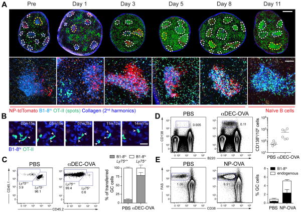

Figures

Figure 1
Figure 1. Expansion kinetics and anatomical distribution of TFH during germinal center formation
(A) 3 × 10⁴ GFP+ OVA-specific T cells (OT-II) and 1.5 × 10⁶ tdTomato+ B1-8hi B cells (corresponding to 1.5 × 10⁵ Igλ+, NP-specific B cells) were transferred into wild type mice 1 d before subcutaneous immunization with NP-OVA in alum. Popliteal LN were explanted at 0, 3, 5, 8, and 11 days after immunization and subjected to whole-lymph node scanning by TPLSM. Collapsed Z-stacks (50–100 μm total volume, 5 μm steps) are shown. Scale bars, 300 μm. Right: quantitation of T cell density and density ratio (T cells in GC/T cells outside of GC) in two independent experiments. (B–C) Expression of TFH markers on PAGFP+ OT-II cells photoactivated in different LN regions as described in Fig. S1. Each symbol represents one experiment.
Figure 2
Figure 2. GC-TFH cells in individual germinal centers are not clonally restricted
(A) A total of 3 × 10⁵ OT-II cells expressing CFP, GFP or DsRed (~105 of each color) and 3–5 × 10⁶ non-fluorescent B1-8hi B cells (corresponding to 3–5 × 10⁵ Igλ+, NP-specific cells) were transferred into WT recipients 1 day before subcutaneous immunization with NP-OVA in alum. Draining LN were explanted 10 days later and imaged by TPLSM. FDC networks were labeled by injection of a mixture of NP-tdTomato and NP-YPet (orange). Central image shows a 50 μm-deep collapsed Z-stack (10 μm steps) of a whole LN containing multiple GCs, magnified in the outer panels. Scale bars, 100 μm (large panel); 50 μm (side panels). (B) Quantitation of data as in (A) across multiple LN from different mice. Each bar represents a single GC. Numbers on top of bars indicate the number of T cells counted in each GC. (C) T and B cells as described in (A) were transferred to H-2Aβ-GFP mice to allow visualization of early GCs, and LN were imaged 5 days after immunization with NP-OVA in alum. Image of one GC and quantitation of multiple GCs are shown. Scale bar, 100 μm. Each set of experiments was performed twice (additional images and quantitation in Fig. S3).
Figure 3
Figure 3. GC-TFH exchange between neighboring germinal centers
(A) GCs containing OT-II PAGFP+ T cells were generated as detailed in Fig. S6A, and the LZ of a single GC was photoactivated within a living mouse. Center panel: collapsed Z-stack image of the cortical half (250 μm depth) of a whole-LN scan taken ~20 hours after photoactivation. Outer panels (i–vi): high-magnification images of the regions indicated in the center panel. Panel vii: original photoactivated GC. Scale bars, 300 μm (center panel), 20 μm (panels i–vi), 30 μm (panel vii). Green spots are placed over photoactivated OT-II cells in low-magnification panels to improve visualization. (B) Quantitation of 3 experiments as in (A) (95, 184 and 208 photoactivated cells analyzed in 3 independent experiments). Each color represents one experiment. Average distance covered by photoactivated cells was 334 μm; the furthest 10% of cells traveled on average 790 μm. (C) Single GCs in popliteal lymph nodes containing PAGFP+ OT-II T cells were photoactivated in vivo as in (A), then explanted and dissected as shown in Fig. S6. FACS plots show PD-1 and CXCR5 expression in emigrant GC-TFH cells (pooled data from 2 mice). Data representative of 5 mice from 3 independent experiments.
Figure 4
Figure 4. Newly-activated TFH can enter ongoing germinal center reactions
(A) Kinetics of invasion of ongoing GCs by newly activated T cells as described in figure S8A. Top: montage showing whole-LN TPLSM scans. Collapsed Z-stacks are 90 μm deep (pre and day 1) or 40 μm deep (days 3–11). GCs containing CFP+ B1-8hi B cells (blue) and NP-tdTomato-coated FDCs (red) are circled with dotted lines. Green spots are placed over GFP+ T cells to improve visualization. Background outside LNs was deleted. Bottom: higher-magnification images of GCs circled in yellow in the top panel. Scale bars, 0.5 mm (top), 100 μm (bottom). (B) Time series showing cognate T-B interactions in an invaded GC on day 11 post-boost. Scale bar, 20 μm. (C–D) Recipient mice were treated with αDEC205-OVA or PBS 6 days after NP-OVA boost as outlined in figure S8D. Flow cytometry plots showing proportion of Ly75+/+ and Ly75−/− B1-8hi GC B cells (C) or percentage of plasmablasts among all single cells (D) 3 days after αDEC205-OVA treatment. (E) Flow cytometry plots comparing the proportion of GC cells (Fas+CD38) in mice treated as outlined in figure S8E or receiving PBS instead of the NP-OVA boost at day 0. Mean ± SEM of 2–3 experiments is depicted in the right-most panels.

References

    1. Vinuesa CG, et al. A RING-type ubiquitin ligase family member required to repress follicular helper T cells and autoimmunity. Nature. 2005 May 26;435:452. - PubMed
    1. Allen CD, Okada T, Tang HL, Cyster JG. Imaging of germinal center selection events during affinity maturation. Science. 2007 Jan 26;315:528. - PubMed
    1. Victora GD, et al. Germinal Center Dynamics Revealed by Multiphoton Microscopy with a Photoactivatable Fluorescent Reporter. Cell. 2010 Nov 12;143:592. - PMC - PubMed
    1. Schwickert TA, et al. A dynamic T cell-limited checkpoint regulates affinity-dependent B cell entry into the germinal center. J Exp Med. 2011 May 16; - PMC - PubMed
    1. Dominguez-Sola D, et al. The proto-oncogene MYC is required for selection in the germinal center and cyclic reentry. Nature immunology. 2012 Nov;13:1083. - PMC - PubMed

Publication types