

Muscarinic receptors as model targets and antitargets for structure-based ligand discovery
- PMID: 23887926
- PMCID: PMC3781386
- DOI: 10.1124/mol.113.087551
Muscarinic receptors as model targets and antitargets for structure-based ligand discovery
Abstract
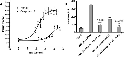
G protein-coupled receptors (GPCRs) regulate virtually all aspects of human physiology and represent an important class of therapeutic drug targets. Many GPCR-targeted drugs resemble endogenous agonists, often resulting in poor selectivity among receptor subtypes and restricted pharmacologic profiles. The muscarinic acetylcholine receptor family exemplifies these problems; thousands of ligands are known, but few are receptor subtype-selective and nearly all are cationic in nature. Using structure-based docking against the M2 and M3 muscarinic receptors, we screened 3.1 million molecules for ligands with new physical properties, chemotypes, and receptor subtype selectivities. Of 19 docking-prioritized molecules tested against the M2 subtype, 11 had substantial activity and 8 represented new chemotypes. Intriguingly, two were uncharged ligands with low micromolar to high nanomolar Ki values, an observation with few precedents among aminergic GPCRs. To exploit a single amino-acid substitution among the binding pockets between the M2 and M3 receptors, we selected molecules predicted by docking to bind to the M3 and but not the M2 receptor. Of 16 molecules tested, 8 bound to the M3 receptor. Whereas selectivity remained modest for most of these, one was a partial agonist at the M3 receptor without measurable M2 agonism. Consistent with this activity, this compound stimulated insulin release from a mouse β-cell line. These results support the ability of structure-based discovery to identify new ligands with unexplored chemotypes and physical properties, leading to new biologic functions, even in an area as heavily explored as muscarinic pharmacology.
Figures

References
-
- Avlani VA, Langmead CJ, Guida E, Wood MD, Tehan BG, Herdon HJ, Watson JM, Sexton PM, Christopoulos A. (2010) Orthosteric and allosteric modes of interaction of novel selective agonists of the M1 muscarinic acetylcholine receptor. Mol Pharmacol 78:94–104 - PubMed
-
- Ballesteros J, Weinstein H. (1995) Integrated methods for modeling G-protein coupled receptors. Methods Neurosci 25:366–428
-
- Blüml K, Mutschler E, Wess J. (1994) Functional role in ligand binding and receptor activation of an asparagine residue present in the sixth transmembrane domain of all muscarinic acetylcholine receptors. J Biol Chem 269:18870–18876 - PubMed
-
- Caccamo A, Fisher A, LaFerla FM. (2009) M1 agonists as a potential disease-modifying therapy for Alzheimer’s disease. Curr Alzheimer Res 6:112–117 - PubMed
Publication types
MeSH terms
Substances
Grants and funding
LinkOut - more resources
Full Text Sources
Other Literature Sources
