. 2013 Sep 15;73(18):5775-86.
doi: 10.1158/0008-5472.CAN-13-1318. Epub 2013 Jul 25.

KIT signaling governs differential sensitivity of mature and primitive CML progenitors to tyrosine kinase inhibitors

Affiliations

KIT signaling governs differential sensitivity of mature and primitive CML progenitors to tyrosine kinase inhibitors

Amie S Corbin et al. Cancer Res. .

Abstract

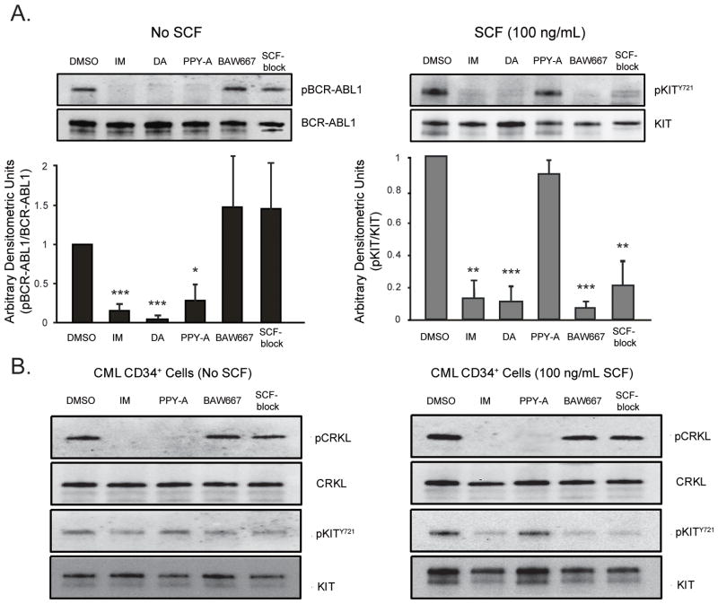
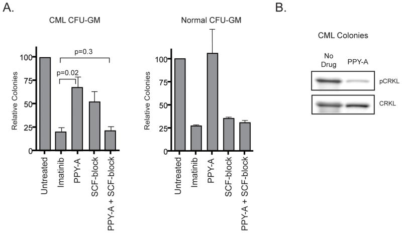
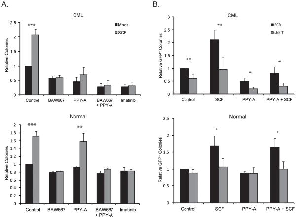
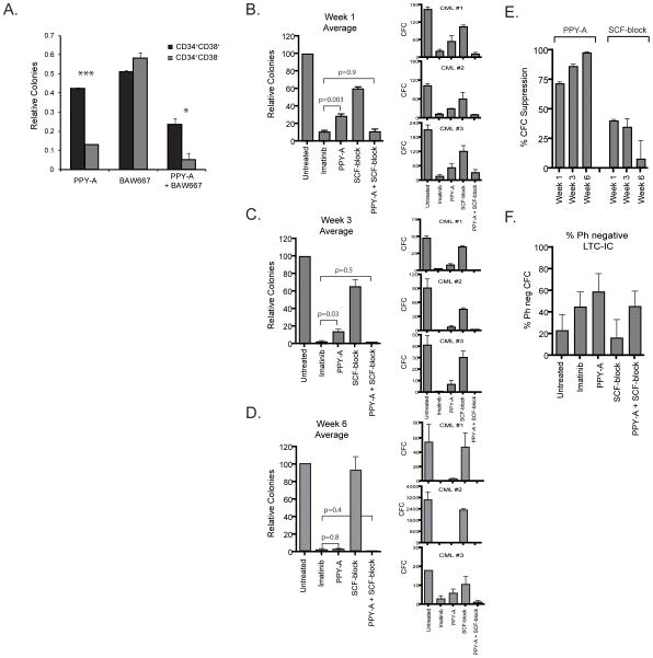
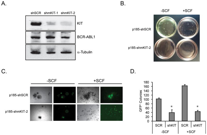
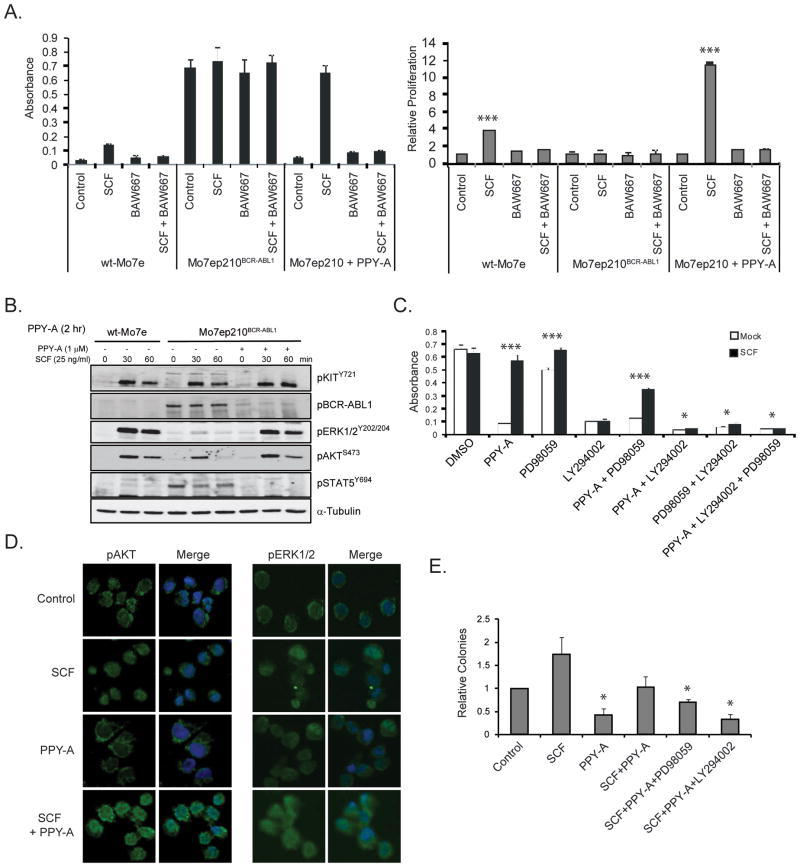
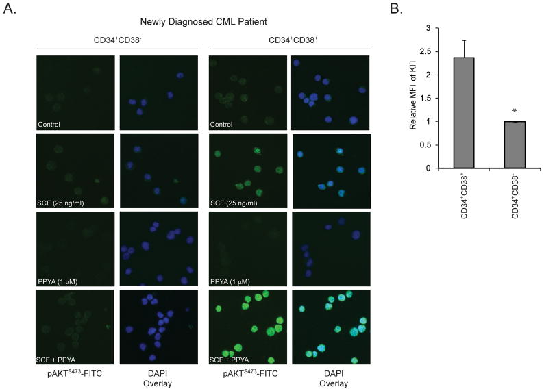
Imatinib and other BCR-ABL1 inhibitors are effective therapies for chronic myelogenous leukemia (CML), but these inhibitors target additional kinases including KIT, raising the question of whether off-target effects contribute to clinical efficacy. On the basis of its involvement in CML pathogenesis, we hypothesized that KIT may govern responses of CML cells to imatinib. To test this, we assessed the growth of primary CML progenitor cells under conditions of sole BCR-ABL1, sole KIT, and dual BCR-ABL1/KIT inhibition. Sole BCR-ABL1 inhibition suppressed mature CML progenitor cells, but these effects were largely abolished by stem cell factor (SCF) and maximal suppression required dual BCR-ABL1/KIT inhibition. In contrast, KIT inhibition did not add to the effects of BCR-ABL1 inhibition in primitive progenitors, represented by CD34(+)38(-) cells. Long-term culture-initiating cell assays on murine stroma revealed profound depletion of primitive CML cells by sole BCR-ABL1 inhibition despite the presence of SCF, suggesting that primitive CML cells are unable to use SCF as a survival factor upon BCR-ABL1 inhibition. In CD34(+)38(+) cells, SCF strongly induced pAKT(S473) in a phosphoinositide 3-kinase (PI3K)-dependent manner, which was further enhanced by inhibition of BCR-ABL1 and associated with increased colony survival. In contrast, pAKT(S473) levels remained low in CD34(+)38(-) cells cultured under the same conditions. Consistent with reduced response to SCF, KIT surface expression was significantly lower on CD34(+)38(-) compared with CD34(+)38(+) CML cells, suggesting a possible mechanism for the differential effects of SCF on mature and primitive CML progenitor cells.


Conflict of interest statement

Disclosure of Potential Conflicts of Interest:

M.W.D. is a consultant for BMS, Novartis, ARIAD and Incyte and his laboratory receives research funding from BMS and Novartis. B.J.D. is principal investigator or co-investigator on Novartis, Bristol-Myers Squibb, and ARIAD clinical trials. His institution has contracts with these companies to pay for patient costs, nurse and data manager salaries, and institutional overhead. He does not derive salary, nor does his lab receive funds from these contracts. OHSU and B.J.D. have a financial interest in MolecularMD. OHSU has licensed technology used in some of these clinical trials to MolecularMD. This potential individual and institutional conflict of interest has been reviewed and managed by OHSU. P.W.M. is an employee of Novartis Pharma, Basel, Switzerland. J.E.C. receives research funding from ARIAD, Novartis, BMS, Pfizer, Chemgenex, Deciphera, and Incyte.

Figures

Figure 1
Figure 1. BCR-ABL1 and KIT inhibitor profile
(A) Mo7ep210BCR-ABL1 or Mo7e cells stimulated with SCF were treated overnight with 2 μM imatinib (IM), 50 nM dasatinib (das), 1 μM BAW667, 1 μM PPY-A or 200 ng/mL SCF blocking antibody (SCF-block). Lysates were immunoblotted for phosphotyrosine or pKITY721 respectively. Total BCR-ABL1 and total KIT are shown as loading controls. Mean relative signal intensity in the presence of inhibiting agents, determined by densitometric quantitation of band intensity, is shown for n=3 replicates. Error bars represent SEM; *p<0.05; **p<0.01; ***p<0.001 (Student’s t-test). (B) CD34+ cells from newly diagnosed chronic phase CML patients (n=3) were treated for 4 hours with 2 μM IM, 1 μM BAW667, 1 μM PPY-A, or 200 ng/mL SCF-block ± SCF stimulation as indicated. Lysates were immunoblotted for pCRKL or pKITY721. Total CRKL and total KIT are shown as loading controls. One representative experiment is shown. Dasatinib was not tested due to cell number limitations.
Figure 2
Figure 2. Suppression of CML and normal CFU-GM colony formation by PPY-A, imatinib, SCF-block or PPY-A+SCF-block
(A) CFU-GM were assessed in samples from newly diagnosed chronic phase CML patient cells (left) or normal mononuclear cells (right) cultured for 14 days in semisolid medium containing IL-3, GM-CSF and SCF. Imatinib, PPY-A, SCF-block or PPY-A+SCF-block were added as indicated. Mean colony number of triplicate plates is shown normalized relative to untreated for n=4 samples. Error bars represent SEM. (B) CFU-GM colonies from untreated and PPY-A-treated plates were pooled and lysates were immunoblotted for pCRKL to assess BCR-ABL1 activity (n=3). Total CRKL is shown as a loading control. A representative immunoblot is shown.
Figure 3
Figure 3. Effects of genetic or biochemical KIT inhibition on PPY-A sensitivity in normal and CML CD34+ cells
(A) CD34+ cells from newly diagnosed CML-CP patients (top, n=4) or healthy controls (bottom, n=4) were cultured for 14 days in semisolid medium containing IL-3 and GM-CSF ± SCF. BAW667, PPY-A, BAW667+PPY-A or imatinib were added as indicated. CFU-GM colonies were scored on day 14. Untreated controls (without SCF) were set to 1. Error bars represent SEM; *p<0.05; **p<0.01; ***p<0.001 (Student’s t-test). (B) CD34+ cells from the same patients (top) and controls (bottom) as in (A) were infected with lentivirus for simultaneous expression of GFP and either shKIT or shSCR. Cells were plated in semisolid medium containing IL-3 and GM-CSF. PPY-A, SCF or both were added as indicated. GFP-positive colonies were scored after 14 days. shSCR controls cultured with IL-3 and GM-SCF only were set to 1. Error bars represent SEM; *p<0.05; **p<0.01; ***p<0.001 (Student’s t-test).
Figure 4
Figure 4. Suppression of mature vs. primitive CML progenitors and stem cells by sole inhibition of BCR-ABL1, sole inhibition of KIT or combined inhibition of KIT and BCR-ABL1
(A) LinCD34+38+ and LinCD34+38 cells were isolated from viably frozen MNC from CML-CP patients (n=3) and plated in semisolid media containing IL-3 and GM-CSF with or without 1 μM PPY-A, 1 μM BAW667 or their combination. CFU-GM colonies were counted after 14 days. Untreated controls were set to 1. Error bars represent SEM. *p<0.05; ***p<0.001 (Student’s t-test). (B–E) A separate series of experiments was performed on CD34+ cells from newly diagnosed CML patients (n=3). Cells were cultured in LTC-IC assays on murine stromal cells (M2-10B4) in the presence of 2 μM imatinib, 1 μM PPY-A, 200 ng/mL SCF-block or PPY-A+SCF-block for a total duration of six weeks. At (B) 1 week, (C) 3 weeks and (D) 6 weeks, triplicate cultures were assessed for CFC growth. Total CFC derived from 10,000 input cells are shown for each sample as well as normalized mean values for each time point. Error bars represent standard error of the mean (SEM). Differences in LTC-IC numbers were evaluated by Student’s t-test. The frequency of Ph+ colonies was determined by FISH analysis of individual colonies from all treatment conditions. Data presented in parts B–C include only Ph+ colonies. (E) Suppression of mature and primitive CML cell outgrowth in the presence of sole BCR-ABL1 or sole KIT inhibition was compared for the three LTC-IC assays combined. (F) Frequency of normal (Ph) colonies in 6-week CML LTC-IC. Mean values for the three samples are shown. Error bars represent SEM.
Figure 5
Figure 5. Effects of KIT knockdown on colony formation by murine bone marrow cells in the presence and absence of SCF
(A) Murine bone marrow MNCs were infected with MIG-p185-SCR, or MIG-p185-shmKIT-2 lentivirus. GFP+ cells were sorted and p185-BCR-ABL1 and KIT expression were detected by immunoblot. (B–D) BM from 5-FU treated mice was infected with MIG-p185-SCR or MIG-p185-shmKIT-2 lentivirus and colony assays were set up in the presence or absence of 50 ng/ml rmSCF without any other cytokines. After 15 days plates were (B) photographed, (C) GFP-positive colonies were identified and counted under a fluorescence microscope and (D) colony numbers were compared between conditions. The experiment was performed twice, with similar results. Error bars represent SEM. *p<0.05 (Student’s t-test).
Figure 6
Figure 6. Effects of SCF in combination with inhibition of BCR-ABL1, KIT, PI3K and/or MEK on Mo7ep210BCR-ABL1, Mo7e cells and primary CD34+ CML-CP cells
(A) Mo7ep210BCR-ABL1 and Mo7e cells were treated with 25 ng/ml SCF, 1 μM PPY-A, 1 μM BAW667 or their combination(s) as indicated. Viable cell numbers were measured after 72 hours. Results at 24 hours and 48 hours were comparable (not shown). Error bars represent SEM. ***p<0.001 (Student’s t-test). (B) Total cellular lysates were harvested after 30 and 60 minutes and subjected to immunoblot analysis for pKITY721, pABLY402, pERK1/2Y202/204, pAKTS473, pSTAT5Y694 and α-Tubulin (loading control). (C) Mo7ep210BCR-ABL1 cells were treated with PPY-A in combination with 20 μM PD98059 (MEK inhibitor) or 20 μM LY294002 (PI3K inhibitor), in the presence or absence of 25 ng/mL SCF. Viable cells were measured by MTS assay at 72 hours. Error bars represent SEM. *p<0.05; ***p<0.001 (Student’s t-test). (D, E) CD34+ cells from newly diagnosed CML-CP patients (n=3) were incubated with or without PPY-A (1 3M) for 2 hours, followed by SCF (25 ng/mL) for 30 minutes. Aliquots of cells were analyzed for pAKTS473 and pERK1/2Y202/204 by immunofluorescence (D) or cultured in semisolid medium, using identical conditions, with CFU-GM colonies assessed after 15 days (E). Error bars represent SEM.*p<0.05 (Student’s t-test).
Figure 7
Figure 7. Assessment of SCF-induced pAKTS473 and CD117 expression in mature and primitive CML progenitor cells
(A) LinCD34+38+ and LinCD34+38 cells from newly diagnosed CML-CP patients (n=3) were simulated with 25 ng/mL SCF with or without prior treatment with 1 μM PPY-A, followed by immunofluorescence to detect pAKTS473. (B) Cryopreserved column-selected CD34+ cells from newly diagnosed CML-CP patients (n=4) were labeled with CD117-PerCP-Cy5.5, Lin-FITC, CD34-APC and CD38-PE antibodies and mean fluorescence intensity of CD117 measured. Error bars represent SEM. *p<0.05 (Student’s t-test).

References

    1. Druker BJ, Guilhot F, O’Brien SG, Gathmann I, Kantarjian H, Gattermann N, et al. Five-year follow-up of patients receiving imatinib for chronic myeloid leukemia. N Engl J Med. 2006;355:2408–17. - PubMed
    1. Deininger M, Buchdunger E, Druker BJ. The development of imatinib as a therapeutic agent for chronic myeloid leukemia. Blood. 2005;105:2640–53. - PubMed
    1. Dewar AL, Cambareri AC, Zannettino AC, Miller BL, Doherty KV, Hughes TP, et al. Macrophage colony-stimulating factor receptor c-fms is a novel target of imatinib. Blood. 2005;105:3127–32. - PubMed
    1. Manley PW, Stiefl N, Cowan-Jacob SW, Kaufman S, Mestan J, Wartmann M, et al. Structural resemblances and comparisons of the relative pharmacological properties of imatinib and nilotinib. Bioorg Med Chem. 2010;18:6977–86. - PubMed
    1. Al-Ali HK, Heinrich MC, Lange T, Krahl R, Mueller M, Muller C, et al. High incidence of BCR-ABL kinase domain mutations and absence of mutations of the PDGFR and KIT activation loops in CML patients with secondary resistance to imatinib. Hematol J. 2004;5:55–60. - PubMed

Publication types

MeSH terms