. 2013 Aug 27;52(34):5760-9.
doi: 10.1021/bi400405p. Epub 2013 Aug 16.

Membrane peroxidation and methemoglobin formation are both necessary for band 3 clustering: mechanistic insights into human erythrocyte senescence

Affiliations

Membrane peroxidation and methemoglobin formation are both necessary for band 3 clustering: mechanistic insights into human erythrocyte senescence

Nobuto Arashiki et al. Biochemistry. .

Abstract

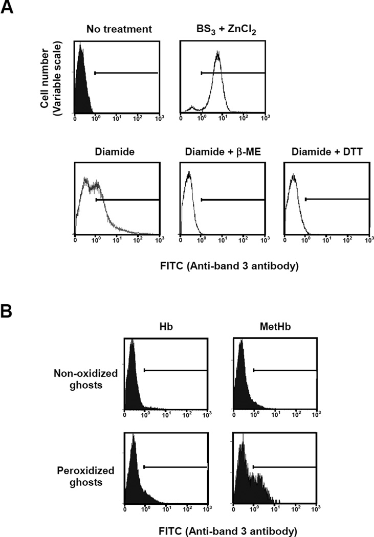
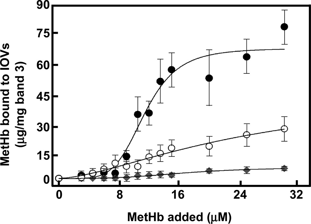
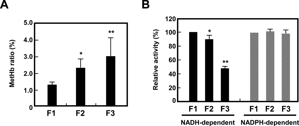
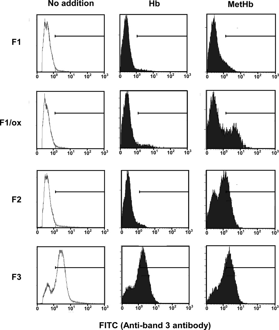
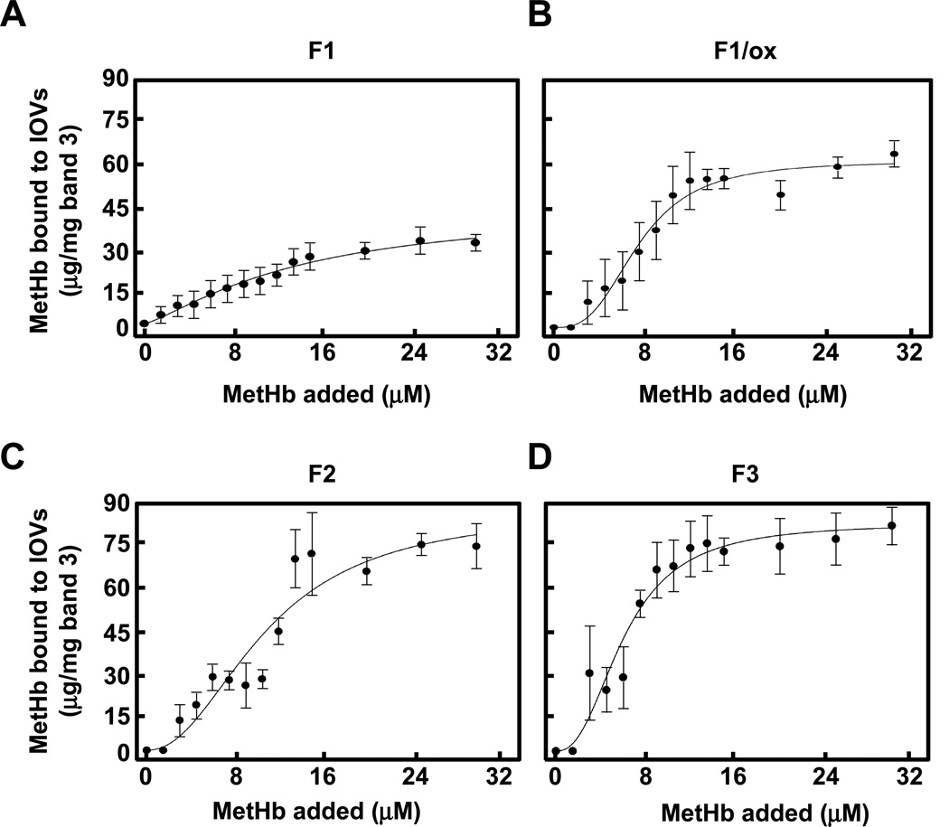
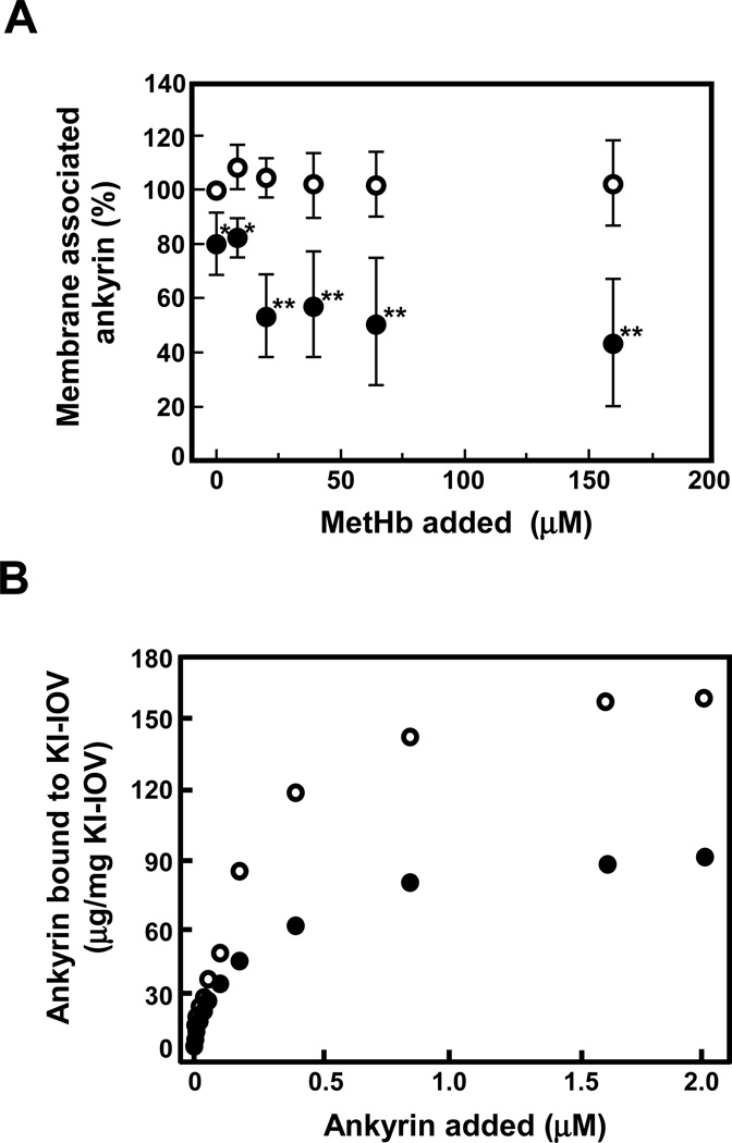
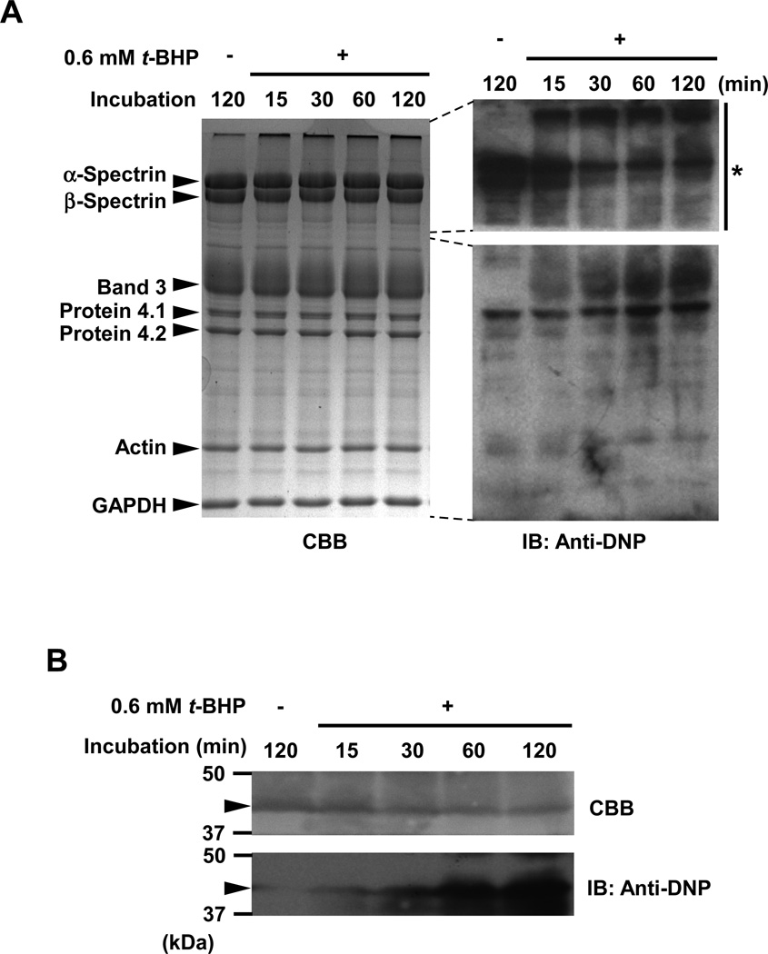
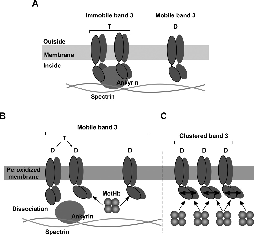
Oxidative damage and clustering of band 3 in the membrane have been implicated in the removal of senescent human erythrocytes from the circulation at the end of their 120 day life span. However, the biochemical and mechanistic events leading to band 3 cluster formation have yet to be fully defined. Here we show that while neither membrane peroxidation nor methemoglobin (MetHb) formation on their own can induce band 3 clustering in the human erythrocytes, they can do so when acting in combination. We further show that binding of MetHb to the cytoplasmic domain of band 3 in peroxidized, but not in untreated, erythrocyte membranes induces cluster formation. Age-fractionated populations of erythrocytes from normal human blood, obtained by a density gradient procedure, have allowed us to examine a subpopulation, highly enriched in senescent cells. We have found that band 3 clustering is a feature of only this small fraction, amounting to ∼0.1% of total circulating erythrocytes. These senescent cells are characterized by an increased proportion of MetHb as a result of reduced nicotinamide adenine dinucleotide-dependent reductase activity and accumulated oxidative membrane damage. These findings have allowed us to establish that the combined effects of membrane peroxidation and MetHb formation are necessary for band 3 clustering, and this is a very late event in erythrocyte life. A plausible mechanism for the combined effects of membrane peroxidation and MetHb is proposed, involving high-affinity cooperative binding of MetHb to the cytoplasmic domain of oxidized band 3, probably because of its carbonylation, rather than other forms of oxidative damage. This modification leads to dissociation of ankyrin from band 3, allowing the tetrameric MetHb to cross-link the resulting freely diffusible band 3 dimers, with formation of clusters.


Figures

Figure 1
Figure 1. FACS analysis of band 3 clustering in normal and variously treated ghosts
(A) To assess the specificity of the peptide antibody recognizing clustered band 3, ghosts prepared from normal erythrocytes were incubated in the absence (upper left panel) or presence (upper right panel) of 1 mM BS3 and ZnCl2 to chemically cross-link band 3 or in the presence of 2 mM diamide (lower left panel) to reversibly cross-link band 3 by disulfide bonds. Moreover, diamide-treated ghosts reduced by 20 mM DTT (lower middle panel) or 10 mM β-ME (lower right panel) were also prepared to assess the reversible effect. FACS analysis of the cells was performed following incubation with rabbit anti-band 3 antibody and FITC-conjugated anti-rabbit IgG antibody. (B) Ghosts prepared from normal erythrocytes were incubated with (peroxidized ghosts) or without (non-oxidized ghosts) 0.6 mM t-BHP, a lipid peroxidation reagent, and then resealed with either Hb or MetHb. These resealed ghosts were subjected to FACS analysis as described above.
Figure 2
Figure 2. Binding of MetHb to IOVs prepared from non-oxidized and peroxidized normal erythrocyte membranes
Ten µg of non-oxidized IOVs (○), IOVs peroxidized by 0.6 mM t-BHP (●), and peroxidized IOVs digested by 5 µg/ml trypsin (♦) were incubated with MetHb (0 to 31.3 µM) and then IOVs were collected in the pellets by centrifugation through 8% sucrose cushion followed by SDS-PAGE and immunoblotting with anti-Hbβ monoclonal antibody. The amount of MetHb bound to IOVs was calculated by densitometric analysis. The data are the mean values ± S.D. of three individual experiments and the lines represent the best fit of the data using the Hill equation.
Figure 3
Figure 3. MetHb content and MetHb reductase activity of fractionated erythrocytes
(A) MetHb content of erythrocytes in F1, F2, and F3 fractions from 4 individuals was measured by the cyan methemoglobin method described in Methods. Data indicate the means ± S.D. *P<0.05, **P<0.01 vs. F1. (B) NADH- and NADPH-dependent MetHb reductase activities were measured in F1, F2, and F3 erythrocytes as described in Methods. Enzyme activities (%) in F2 and F3 erythrocytes were expressed as relative to the values derived for F1 erythrocytes. Data indicate the means ± S.D. (n = 3). *P<0.05, **P<0.01 vs. F1.
Figure 4
Figure 4. FACS analysis of Hb- or MetHb-incorporated ghosts prepared from density fractionated erythrocytes
Hb or MetHb were resealed into the ghosts made from erythrocytes isolated in F1, F2, and F3 fractions, and peroxidized F1 ghosts (F1/ox). Band 3 clustering in these resealed ghosts was analyzed by FACS as described in the Methods.
Figure 5
Figure 5. Binding of MetHb to IOVs prepared from density fractionated erythrocytes
The amount of MetHb bound to IOVs prepared from F1 (A), F1/ox (B), F2 (C), and F3 (D) ghosts was measured and showed as described in the Methods and the legend of Fig. 2.
Figure 6
Figure 6. Interaction of ankyrin with band 3 in non-oxidized and peroxidized IOVs
(A) The amount of ankyrin associated with non-oxidized (○) or peroxidized (●) IOVs was measured in the presence or absence of MetHb by immunoblotting using anti-ankyrin antibody as described in Methods. The data represent the mean ratio (%) ± S.D. normalized by the amount of ankyrin bound to non-oxidized IOVs in the absence of MetHb (n = 3). *P<0.05, **P<0.01. (B) KI-IOVs pretreated with (●) or without (○) t-BHP were incubated with increasing concentrations of purified ankyrin. The amount of bound ankyrin to KI-IOVs was measured as described in Method and plotted against the concentration of ankyrin added.
Figure 7
Figure 7. Carbonylation of band 3 induced by membrane peroxidation
Carbonylated proteins of the ghosts which had been treated with or without 0.6 mM t-BHP for up to 120 min were analyzed as described in Methods. (A) Signal intensity of carbonylated proteins corresponding to the position of band 3 in the gel increased in time-dependent manner. The asterisk indicates a part of the gel that was exposed for a very short period (5 sec), showing that spectrin was endogenously carbonylated under the experimental conditions used to monitor carbonylation but did not show time-dependent changes. (B) Carbonylation of cytoplasmic domain of band 3 (43 kDa; arrow head) in t-BHP-treated IOVs. Carbonylation of cytoplasmic domain of band 3 increased with increasing incubation time.
Figure 8
Figure 8. A potential mechanism for the combined effect of membrane peroxidation and MetHb in inducing band 3 clustering
(A) Band 3 tetramer formed by two dimers and linked to the skeletal network through interaction with ankyrin is immobile and is thus uniformly distributed in the membrane. The mobile band 3 dimer is not connected to the skeleton but its area of diffusion is restricted to within the lattice (fence) of the network. (B) Peroxidation of erythrocyte membranes results in a modest degree of dissociation of ankyrin from cytoplasmic domain of band 3 and high affinity cooperative binding of MetHb to cytoplasmic domain of band 3 accelerates this dissociation process probably due to carbonylation of cytoplasmic domain of band 3. Following dissociation from ankyrin, band 3 is released from its constraints for lateral diffusion in the membrane. (C) Binding of MetHb to one subunit of cytoplasmic domain of band 3 induces a conformational change in the cytoplasmic domains of other associated band 3 subunits leading to high affinity cooperative binding of MetHb. As MetHb is a tetramer, it can form a bridge between a number of diffusing band 3 dimers to induce clustering.

References

    1. Burak Cimen MY. Free radical metabolism in human erythrocytes. Clinica. Chimica. Acta. 2008;390:1–11. - PubMed
    1. Pandey KB, Rizvi SI. Markers of oxidative stress in erythrocytes and plasma during aging in humans. Oxid. Med. Cell. Longev. 2010;3:2–12. - PMC - PubMed
    1. Signorini C, Ferrali M, Ciccoli L, Sugherini L, Magnani A, Comporti M. Iron release, membrane protein oxidation and erythrocyte ageing. FEBS Lett. 1995;362:165–170. - PubMed
    1. Hochstein P, Jain SK. Association of lipid peroxidation and polymerization of membrane proteins with erythrocyte aging. Fed. Proc. 1981;40:183–188. - PubMed
    1. Jain SK. Evidence for membrane lipid peroxidation during the in vivo aging of human erythrocytes. Biochim. Biophys. Acta. 1988;937:205–210. - PubMed

Publication types

MeSH terms

Substances

LinkOut - more resources