. 2013 Jul 24;79(2):281-92.
doi: 10.1016/j.neuron.2013.05.015.

The transcription factor Mef2 links the Drosophila core clock to Fas2, neuronal morphology, and circadian behavior

Affiliations

The transcription factor Mef2 links the Drosophila core clock to Fas2, neuronal morphology, and circadian behavior

Anna Sivachenko et al. Neuron. .

Abstract

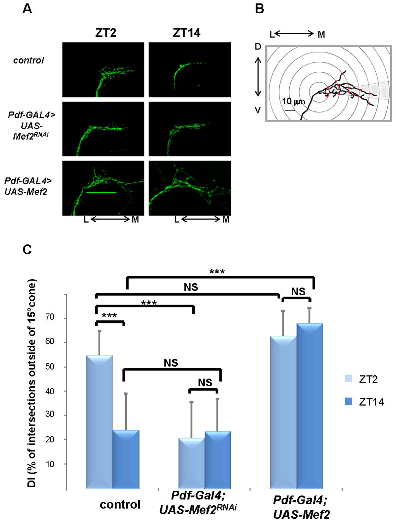
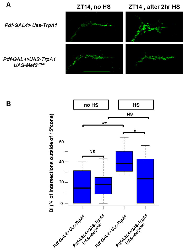
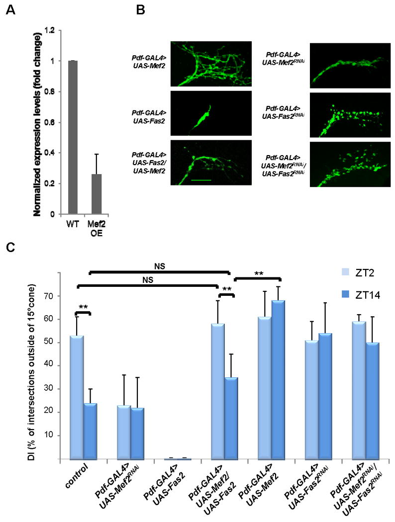
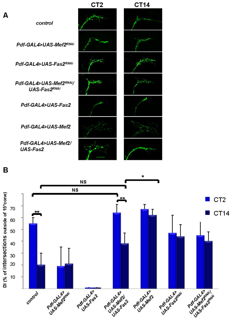
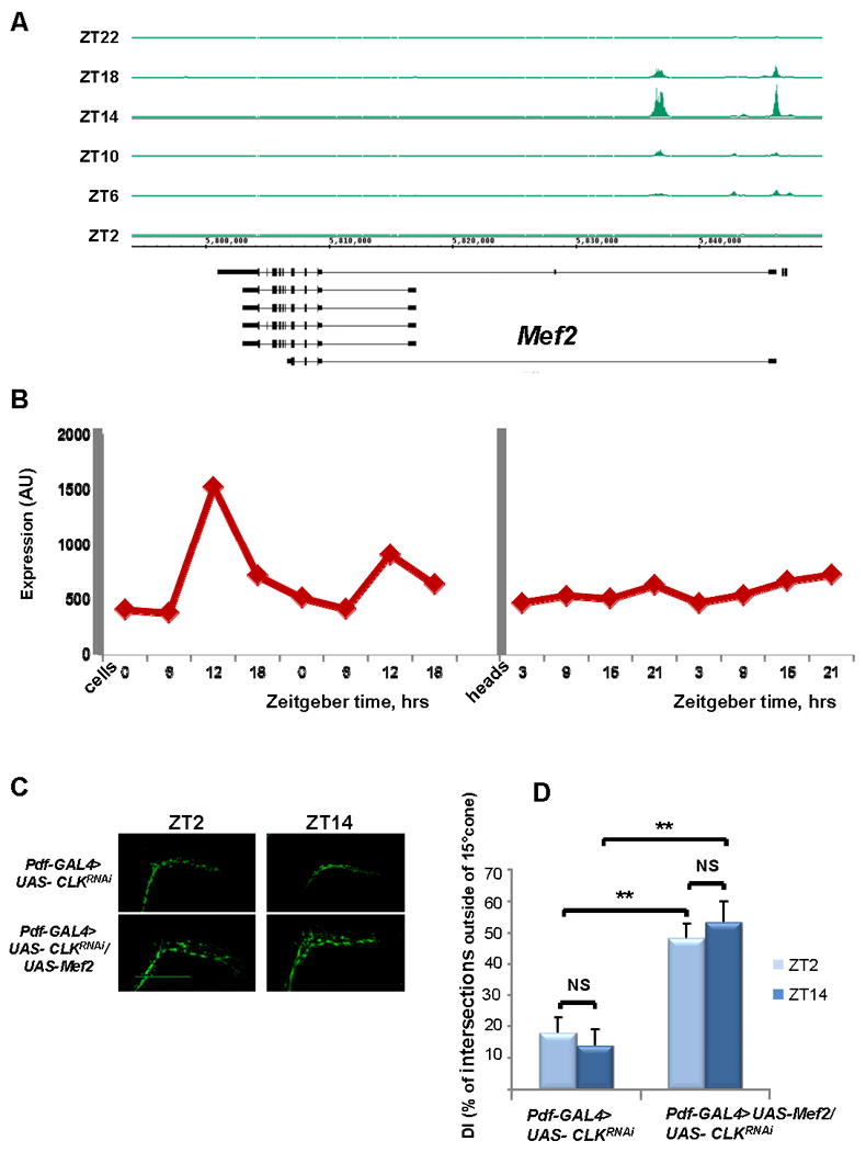
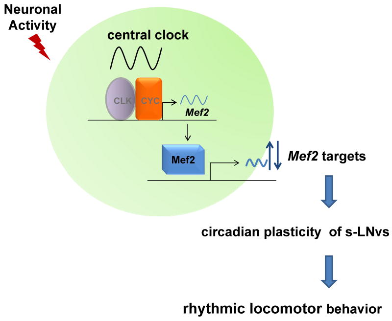
The transcription factor Mef2 regulates activity-dependent neuronal plasticity and morphology in mammals, and clock neurons are reported to experience activity-dependent circadian remodeling in Drosophila. We show here that Mef2 is required for this daily fasciculation-defasciculation cycle. Moreover, the master circadian transcription complex CLK/CYC directly regulates Mef2 transcription. ChIP-Chip analysis identified numerous Mef2 target genes implicated in neuronal plasticity, including the cell-adhesion gene Fas2. Genetic epistasis experiments support this transcriptional regulatory hierarchy, CLK/CYC- > Mef2- > Fas2, indicate that it influences the circadian fasciculation cycle within pacemaker neurons, and suggest that this cycle also contributes to circadian behavior. Mef2 therefore transmits clock information to machinery involved in neuronal remodeling, which contributes to locomotor activity rhythms.


Figures

Figure 1
Figure 1. Genetic manipulations of Mef2 levels disrupt circadian and activity-dependent changes in axonal fasciculation of the s-LNv dorsal projections
A) Representative confocal images of brains of control flies (yw, UAS-mCD8 GFP; Pdf –Gal4), and flies with reduced (yw, UAS-mCD8 GFP; Pdf-Gal4/+; UAS-Mef2RNAi/+) or increased (yw,UAS-mCD8 GFP; Pdf-Gal4/+; UAS-Mef2/+) levels of Mef2 in PDF cells at ZT2 and ZT14. Note open, defasciculated axonal conformation at ZT2 and fasciculated axons at ZT14 in control flies. Mef2RNAi flies exhibit fasciculated axonal morphology at both circadian time points, while Mef2 overexpression leads to severe axonal defasciculation. Scale bar 50μm. B) Schematic representing modified Sholl's analysis for quantification of fasciculation of axonal termini of s-LNv neurons (see Experimental Procedures). C) Analysis of axonal morphology (fasciculation) of s-LNv dorsal termini by modified Sholl's analysis. The plot represents percentage of intersections between concentric rings and axonal branches outside of a 15% cone (defasciculation index, DI). Statistically significant difference in DI between different genotypes and different time points is depicted by brackets. Circadian variation in DI at ZT2 and ZT14 is abolished in the flies with both decreased and increased Mef2 levels. Plots show mean values, error bars are SEM. *** represents p-value < 0.0001 (non parametric Mann-Whitney test). NS: not significant. For each time point and genotype 20 to 30 brains were analyzed. Two independent experiments were performed with similar results. See also Fig. S1.
Figure 2
Figure 2. Mef2 is required for activity-dependent remodeling of s-LNv axonal projections
A) Induction of TrpA1 in PDF cells by 2 hour temperature elevation to 29°C leads to open conformation of s-LNv dorsal projections at ZT14 in a wild type background. This effect is markedly reduced in an UAS-Mef2RNAi genetic background. Confocal images of fly brains after a 2 hour TrpA1 induction are representative of ∼ 90% of yw; Pdf-Gal4, UAS-mCD8GFP /UAS-TrpA1 flies and of ∼60% of yw; Pdf-Gal4, UAS-mCD8GFP /UAS-TrpA1; UAS-Mef2RNAi/+flies. Scale bar 25 μm. B) Quantification of TrpA1- induced changes in axonal fasciculation by modified Sholl's analysis. Box plot diagram of defasciculation indexes (DI) in the same genotypes as in Figure 2A. Statistically significant difference in axonal fasciculation upon TrpA1 induction is observed in wild type (p-value <0.005, non parametric Mann-Whitney test), but not in Mef2 RNAi genetic background (p-value=0.5). Morphology of dorsal termini after TrpA1 activation significantly differs between Pdf-GAL4>UAS-TrpA1 and Pdf-GAL4>UAS-TrpA1+UAS-Mef2RNAi flies (p-value =0.01). ** represents p-value < 0.005, * represents p-value = 0.01 (non parametric Mann-Whitney test). NS: not significant. 16 to 20 brains were analyzed for each genotype and experimental condition. See also Fig. S2.
Figure 3
Figure 3. Mef2 transcriptional target Fas2 affects neuronal morphology and is genetically epistatic to Mef2
A) Fas2 is negatively regulated by Mef2 in Pdf cells. Mef2 overexpression in Pdf cells results in a marked decrease in Fas2 mRNA levels within those cells. RNA was extracted from PDF cells purified from yw; Pdf-Gal4, UAS-mCD8 GFP (wild type control WT) and yw; Pdf-Gal4, UAS-mCD8 GFP /+; UAS-Mef2/+ flies (Mef2 overexpression OE) at ZT12. The mRNA values for Fas2 were normalized to those of RPL32 (see also Fig. S4 and Table S2 for primer sequences). Plots show mean values, error bars are SEM. B) Overexpression of Fas2 in PDF cells leads to collapse and overfasciculation of s-LNv axonal arbor in wild type background, and rescues defasciculated phenotype in Mef2 overexpression background. Pdf-Gal4, UAS-mCD8 GFP>UAS-Fas2RNAi flies exhibit open conformation of s-LNv axons. The same phenotype is observed when UAS-Fas2RNAi is expressed in Pdf-GAL4>UAS-Mef2RNAi background, suggesting that Fas2 is genetically downstream and possibly is negatively regulated by Mef2. Images are taken at ZT2. Scale bar 25 μm. C) Analysis of axonal morphology (fasciculation) of s-LNv dorsal termini by modified Sholl's analysis at ZT2 and ZT14 in LD in the same genotypes as in Fig. 3B. Pdf-GAL4>UAS-Fas2 flies display severely overfasciculated conformation at both ZT2 and ZT14. Dorsal s-LNv projections undergo normal circadian remodeling in Pdf-GAL4>UAS-Fas2/UAS-Mef2 flies. Pdf-GAL4>UAS- Fas2RNAi and Pdf-GAL4>UAS- Fas2RNAi/UAS- Mef2RNAi show similar defasciculated phenotype and no significant differences in DI between ZT2 and ZT14. Plots show mean values, error bars are SEM. ** represents Pvalue <0.01, non-parametric Mann-Whitney test. NS: not significant. See also Fig. S5.
Figure 4
Figure 4. PDF-cell specific increases in Fas2 levels rescue abnormal axonal morphology and circadian plasticity in Mef2 overexpression background in DD
A) Representative confocal images of yw; Pdf-GAL4, UAS-mCD8 GFP (control), yw; Pdf-GAL4, UAS-mCD8 GFP/+; UAS-Mef2RNAi/+, yw; Pdf-GAL4, UAS-mCD8 GFP >UAS- Fas2RNAi, yw; Pdf-GAL4, UAS-mCD8 GFP >UAS- Fas2RNAi/UAS- Mef2RNAi, yw; Pdf-GAL4, UAS-mCD8 GFP/+; UAS-Fas2 /+, yw; Pdf-GAL4, UAS-mCD8 GFP/+; UAS-Mef2/+, and yw; Pdf-GAL4, UAS-mCD8 GFP/+;UAS-Mef2/UAS-Fas2 fly brains at CT2 and CT14 on the day 2 in DD. Scale bar 25 μm. B) Quantitative analysis of axonal morphology in the same genotypes as in Fig. 4A on day 2 in DD. Overexpression of Fas2 in Pdf-Gal4>UAS-Mef2 background increases axonal fasciculation at CT14 and restores circadian variations in DI. No significant difference between Pdf-Gal4>UAS-Mef2/UAS-Fas2 and control flies observed at both CT2 and CT14. 10 - 12 brains were analyzed for each genotype and time point, experiment was performed twice with very similar results. Plots show mean values, error bars are SEM. ** represents p-value < 0.01, * represents p-value =0.05, non parametric Mann-Whitney test. NS: not significant. See also Fig. S5.
Figure 5
Figure 5. Mef2 is a direct target of CLK/CYC
A) Cyclical CLK binding to Mef2 promoter region. CLK ChIP was performed at 6 time points (ZT2, ZT6, ZT10, ZT14, ZT18, and ZT22), and immunoprecipitated DNA was analyzed by tiling arrays (Affymetrix). CLK binding is visualized with Integrated Genome Browser (IGB). Mef2 is on the bottom strand and therefore transcription occurs from right to left. CLK binds to the 5′-end of Mef2 and binding is maximal at ZT14. B) Mef2 mRNAs cycles in large LNv pacemaker cells but not in heads. Mef2 RNA from either heads(McDonald and Rosbash, 2001) or LNvs(Kula-Eversole et al., 2010; Nagoshi et al., 2010) was analyzed via expression microarrays (Affymetrix). Levels of Mef2 mRNA are shown across four timepoints in clock neurons (ZT0, ZT6, ZT12, and ZT18) and in heads (ZT3, ZT9, ZT15, and ZT21). (C,D) Mef2 expression in PDF cells rescues Clk RNAi-induced increase in axonal fasciculation. Reduction of Clk levels in PDF cells by RNAi results in loss of the circadian plasticity and increased fasciculation of s-LNv dorsal projections. Concurrent overexpression of Mef2 in PDF cells leads to defasciculated axonal conformation resembling a Mef2 overexpression phenotype. (C) Representative confocal images of yw; Pdf-GAL4, UAS-mCD8 GFP/+; UAS- ClkRNAi/+ and yw; Pdf-GAL4, UAS- mCD8 GFP/+; UAS- ClkRNAi/UAS-Mef2 fly brains at ZT2 and ZT14. Scale bar 50 μm. (D) Quantification of axonal fasciculation of dorsal projections of s-LNvs in the same genotypes as in Figure 5C. Modified Sholl's analysis reveals increased fasciculation and lack of circadian variation in the defasciculation index (DI) in yw; Pdf-GAL4, UAS- mCD8 GFP/+; UAS- ClkRNAi/+ axons (p-value =0.22). Co-expression of UAS-Mef2 transgene results in statistically significant increased DI as compared to ClkRNAi (P value < 0.005 at both ZT2 and ZT14), but doesn't rescue the circadian plasticity of s-LNv axons (p-value = 0.16). Plots show mean values, error bars are SEM. ** represents p-value < 0.005, non parametric Mann-Whitney test. NS: not significant. At least 10 brains were analyzed for each genotype and time point. The experiment was performed twice with very similar results.
Figure 6
Figure 6. Mef2 integrates the core circadian oscillator with neuronal activity to regulate neuronal morphology in s-LNv neurons
Rhythmic binding of CLK/CYC to Mef2 promoter results in the oscillations of the Mef2 transcript, as well as cycling of Mef2 protein levels in PDF cells. Mef2 then directly regulates a large group of genes that function in neuronal remodeling, such as Fas2, thus linking the core molecular clock to morphological changes in s-LNv projections. Neuronal activity may possibly influence s-LNv remodeling by modulating the core molecular clock, Mef2 transcriptional activity, or directly affecting post-transcriptional regulation or function of Mef2 target gene products. Rhythmic changes in s-LNv neuronal morphology can serve as a mechanism by which core circadian neurons transmits clock information to downstream systems, which ultimately results in rhythmic locomotor activity.

References

    1. Abruzzi KC, Rodriguez J, Menet JS, Desrochers J, Zadina A, Luo W, Tkachev S, Rosbash M. Drosophila CLOCK target gene characterization: implications for circadian tissue-specific gene expression. Genes Dev. 2011;25:2374–2386. - PMC - PubMed
    1. Appelbaum L, Wang G, Yokogawa T, Skariah GM, Smith SJ, Mourrain P, Mignot E. Circadian and homeostatic regulation of structural synaptic plasticity in hypocretin neurons. Neuron. 2010;68:87–98. - PMC - PubMed
    1. Barbosa AC, Kim MS, Ertunc M, Adachi M, Nelson ED, McAnally J, Richardson JA, Kavalali ET, Monteggia LM, Bassel-Duby R, et al. MEF2C, a transcription factor that facilitates learning and memory by negative regulation of synapse numbers and function. Proc Natl Acad Sci U S A. 2008;105:9391–9396. - PMC - PubMed
    1. Behrens UD, Wagner HJ. Adaptation-dependent changes of bipolar cell terminals in fish retina: effects on overall morphology and spinule formation in Ma and Mb cells. Vision Res. 1996;36:3901–3911. - PubMed
    1. Black BL, Molkentin JD, Olson EN. Multiple roles for the MyoD basic region in transmission of transcriptional activation signals and interaction with MEF2. Mol Cell Biol. 1998;18:69–77. - PMC - PubMed

MeSH terms

Associated data