. 2013 Jul 27:13:194.
doi: 10.1186/1472-6882-13-194.

Neutral sphingomyelinase 2 modulates cytotoxic effects of protopanaxadiol on different human cancer cells

Affiliations

Neutral sphingomyelinase 2 modulates cytotoxic effects of protopanaxadiol on different human cancer cells

Bonggoo Park et al. BMC Complement Altern Med. .

Abstract

Background: Some of ginsenosides, root extracts from Panax ginseng, exert cytotoxicity against cancer cells through disruption of membrane subdomains called lipid rafts. Protopanaxadiol (PPD) exhibits the highest cytotoxic effect among 8 ginsenosides which we evaluated for anti-cancer activity. We investigated if PPD disrupts lipid rafts in its cytotoxic effects and also the possible mechanisms.

Methods: Eight ginsenosides were evaluated using different cancer cells and cell viability assays. The potent ginsenoside, PPD was investigated for its roles in lipid raft disruption and downstream pathways to apoptosis of cancer cells. Anti-cancer effects of PPD was also investigated in vivo using mouse xenograft model.

Results: PPD consistently exerts its potent cytotoxicity in 2 cell survival assays using 5 different cancer cell lines. PPD disrupts lipid rafts in different ways from methyl-β-cyclodextrin (MβCD) depleting cholesterol out of the subdomains, since lipid raft proteins were differentially modulated by the saponin. During disruption of lipid rafts, PPD activated neutral sphingomyelinase 2 (nSMase 2) hydrolyzing membrane sphingomyelins into pro-apoptotic intracellular ceramides. Furthermore, PPD demonstrated its anti-cancer activities against K562 tumor cells in mouse xenograft model, confirming its potential as an adjunct or chemotherapeutic agent by itself in vivo.

Conclusions: This study demonstrates that neutral sphingomyelinase 2 is responsible for the cytotoxicity of PPD through production of apoptotic ceramides from membrane sphingomyelins. Thus neutral sphingomyelinase 2 and its relevant mechanisms may potentially be employed in cancer chemotherapies.


Figures

Figure 1
Figure 1
Protopanaxadiol (PPD) exhibited the most potent cytotoxic effects on cancer cells of various malignancies. K562 cells were treated with PPD at 0, 25 or 50 μM for 24 or 48 hr, then their viabilities were assessed using XTT (A) and SRB assays (B). *, p < 0.05; **, p < 0.01; ***, p < 0.001 for control (vehicle) are considered as significant.
Figure 2
Figure 2
A variety of cancer cells showed high to moderate sensitivities to PPD treatment. (A) Structure of PPD. (B) GI50’s of PPD was calculated against 13 cancer cell lines. GI50 is the concentration that inhibits cell growth by 50%.
Figure 3
Figure 3
Effects of PPD on cell cycle progression of cancer cells were analyzed using flow cytometry. (A) K562 cells were exposed to PPD at 0, 12.5, 25 and 50 μM for 48 hr. Then, they were fixed with ethanol, stained with propidium iodide and analyzed using flow cytometry. (B) K562 cells were exposed to PPD at 0, 12.5 and 25 μM for 24 hr, then expression levels of cell cycle regulators and pro-apoptotic proteins were determined with Western blotting using anti-cdk2, -cdk4, -cdk6, -cyclinA and -cyclinB1 antibodies, -PARP, -Bcl-2, and -caspase-9 antibodies. (C) Relative percentages of cells were calculated at sub-G1, G1, S and G2/M phases. (D) All experiments were repeated three times with similar results, so the means ± standard deviations of band intensities were calculated in our Western blot analyses for cell cycle and apoptosis components. *, p < 0.05; **, p < 0.01; ***, p < 0.001 for control (vehicle) are considered as significant.
Figure 4
Figure 4
PPD disrupted lipid rafts in plasma membranes of cancer cells through cell type specific mechanisms. (A) K562 and HT29 cells were exposed to PPD at 0, 25 and 50 μM in presence of 0, 0.5, 1.0 or 2.5 mg/ml MβCD for 24 hr. The cell viabilities were measured using WST-1 assay. (B) K562 and HT29 cells were exposed to MβCD at 0, 1.0 or 2.5 mg/ml and PPD at 0, 25 or 50 μM for 24 hr, then expression levels of lipid raft-associated proteins were determined with Western blotting using anti-IGF-1R, -pAkt, -caspase-8, -PARP, and -Bid antibodies. (C) All experiments were repeated three times with similar results, so the means ± standard deviations of band intensities were calculated in our Western blot analyses for lipid raft-associated proteins. *, p < 0.05; **, p < 0.01; ***, p < 0.001 for control (vehicle) are considered as significant.
Figure 5
Figure 5
PPD exerted its cytotoxic effects on cancer cells through activation of neutral sphingomyelinase 2. (A) K562 cells were exposed to PPD at 0 (DMSO) and 25 μM for 24 hr. They were stained with BODIPY-Cholesterol ester or BODIPY-C12-SM, then examined using confocal microscopy. (B) K562 cells were exposed to PPD at 0, 12.5, 25 and 50 μM for 6 or 16 hr, then levels of different ceramides were analyzed using LC-MS/MS. K562 (C) and HT29 (D) cells were preincubated with neutral sphingomyelinase (nSMase) inhibitor GW4869 for 1 hr, exposed to PPD at 0, 25 and 50 μM for 48 hr and evaluated for cell viabilities using WST-1 assay. Inhibition studies with GW4869 were repeated three times with similar results. K562 (E) and HT29 (F) cells were transfected with nSMase 2 siRNA 1141725, exposed to PPD at 0, 6.25, 12.5, 25 and 50 μM for 48 hr and evaluated for cell viabilities using WST-1 assay. Inhibition studies with siRNA transfections were repeated twice with similar results. *, p < 0.05; **, p < 0.01; ***, p < 0.001 for control (vehicle) are considered as significant.
Figure 6
Figure 6
PPD sensitized cancer cells to doxorubicin treatment. K562 (A and B) and HT29 (C and D) cells were exposed to Cisplatin (A and C) or PPD (B and D) at 0, 25 and 50 μM in presence of 0, 0.25, 1.0 or 2.5 μM Doxorubicin for 48 hr. All experiments were repeated three times with similar results.
Figure 7
Figure 7
Sensitization by PPD is mediated through altering cell cycle progression and apoptosis. K562 cells were exposed to Cisplatin (A and B) or PPD (C and D) at 0, 25 or 50 μM with or without 0.25 μM Doxorubicin for 48 hr. Then they were fixed with ethanol, stained with propidium iodide and analyzed using flow cytometry. All experiments were repeated three times with similar results.
Figure 8
Figure 8
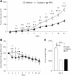
PPD reduced both tumor volumes and weights in in vivo xenograft mouse model. K562-xenografted BALB/c nude mice were randomly divided and treated daily with vehicle, Cisplatin (5 mg/kg) or PPD (25 or 50 mg/kg), when their tumor volumes reached 47.6 mm3. (A) The tumor volumes were determined using the formula (width2 × length)/2, where width represents the smaller diameter. (B) The body weights were measured daily during treatments. Body weight changes were given as percentage of mouse body weights on day 0. (C) The tumor weights were measured on the final 11th day. Unpaired Student’s t test was used for comparisons between control (vehicle) and Cisplatin or PPD group. *, p < 0.05; **, p < 0.01; ***, p < 0.001 for control (vehicle) and Cisplatin, and †, p < 0.05; ††, p < 0.01; †††, p < 0.001 for control (vehicle) and PPD are considered as significant.

Similar articles

Cited by

References

    1. Leung KW, Wong AS. Pharmacology of ginsenosides: a literature review. Chin Med. 2010;5:20. doi: 10.1186/1749-8546-5-20. - DOI - PMC - PubMed
    1. Nag SA, Qin JJ, Wang W, Wang MH, Wang H, Zhang R. Ginsenosides as anticancer agents: In vitro and in vivo activities, structure-activity relationships, and molecular mechanisms of action. Front Pharmacol. 2012;3:25. - PMC - PubMed
    1. Wang W, Zhao Y, Rayburn ER, Hill DL, Wang H, Zhang R. In vitro anti-cancer activity and structure-activity relationships of natural products isolated from fruits of Panax ginseng. Cancer Chemother Pharmacol. 2007;59:589–601. doi: 10.1007/s00280-006-0300-z. - DOI - PubMed
    1. Babina IS, Donatello S, Nabi IR, Hopkins AM. In: Breast cancer - carcinogenesis, cell growth and signalling pathways. Gunduz M, Gunduz E, editor. New York: INTECH; 2011. Lipid Rafts as Master Regulators of Breast Cancer Cell Function; pp. 401–428.
    1. George KS, Wu S. Lipid raft: a floating island of death or survival. Toxicol Appl Pharmacol. 2012;259:311–319. doi: 10.1016/j.taap.2012.01.007. - DOI - PMC - PubMed

Publication types