. 2013 Jul 25;20(7):912-21.
doi: 10.1016/j.chembiol.2013.05.010.

PIKfyve, a class III PI kinase, is the target of the small molecular IL-12/IL-23 inhibitor apilimod and a player in Toll-like receptor signaling

Affiliations

PIKfyve, a class III PI kinase, is the target of the small molecular IL-12/IL-23 inhibitor apilimod and a player in Toll-like receptor signaling

Xinming Cai et al. Chem Biol. .

Abstract

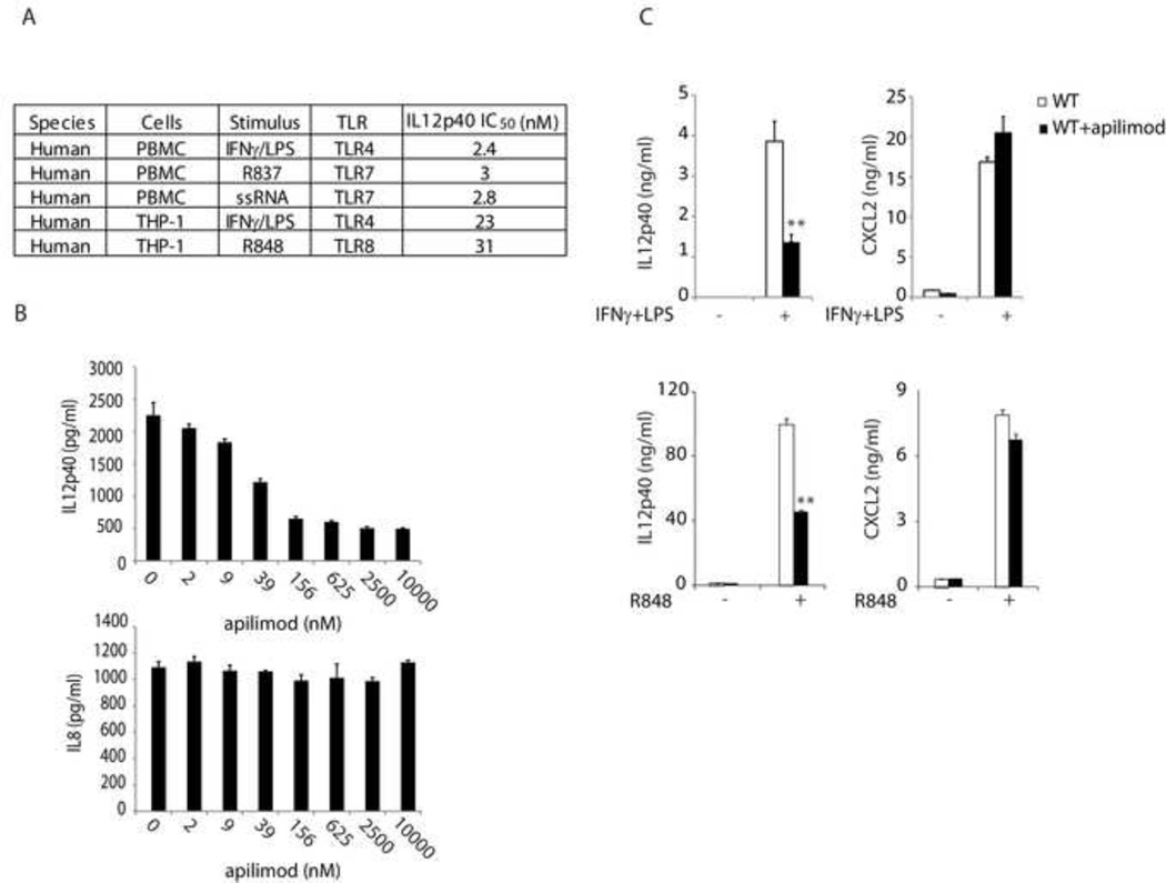
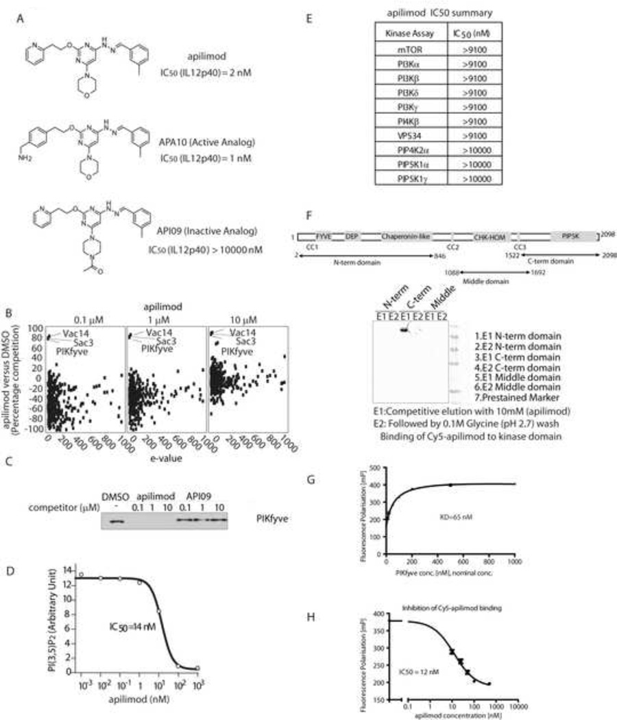
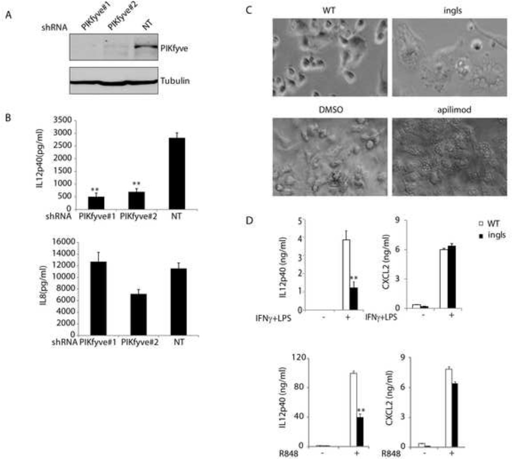
Toll-like receptor (TLR) signaling is a key component of innate immunity. Aberrant TLR activation leads to immune disorders via dysregulation of cytokine production, such as IL-12/IL-23. Herein, we identify and characterize PIKfyve, a lipid kinase, as a critical player in TLR signaling using apilimod as an affinity tool. Apilimod is a potent small molecular inhibitor of IL-12/IL-23 with an unknown target and has been evaluated in clinical trials for patients with Crohn's disease or rheumatoid arthritis. Using a chemical genetic approach, we show that it binds to PIKfyve and blocks its phosphotransferase activity, leading to selective inhibition of IL-12/IL-23p40. Pharmacological or genetic inactivation of PIKfyve is necessary and sufficient for suppression of IL-12/IL-23p40 expression. Thus, we have uncovered a phosphoinositide-mediated regulatory mechanism that controls TLR signaling.


Figures

Figure 1
Figure 1. Apilimod selectively inhibits TLR-induced cytokine expression
The production of cytokines was measured by ELISA following overnight stimulation. Representative results were shown from three independent experiments. (A) Inhibition of IL12p40 by apilimod following stimulation of cells with IFNγ (50 ng/ml)/LPS (1 µg/ml), R837 (10 µg/ml) and R848 (10 µg/ml), or ssRNA (ORN 02, 5 µg/ml). (see also Figure S1) (B) THP-1 cells were treated with apilimod in the presence of IFNγ (50 ng/ml)/LPS (1 µg/ml). The data were analyzed using one-way Anova method (P<0.0001), indicating a significant effect of apilimod on TLR4-induced expression of IL12p40. (C) Mouse BMDCs were treated with apilimod (1 µM) and challenged with IFNγ (50 ng/ml)/LPS (1 µg/ml) or R848 (0.1 µM). **, P<0.01 using Student’s t-Test. Data represent mean values ± SD.
Figure 2
Figure 2. Apilimod binds to and inhibits PIKfyve kinase activity
(A) Structure of apilimod and its analogs and their IC50 for IFNγ (50 ng/ml)/LPS (1 µg/ml) induced-IL12p40 secretion in THP-1 cells. (B) Scatter plot depicting proteins identified in a quantitative chemical proteomics experiment. Proteins are plotted as a function of the percentage competition with apilimod relative to DMSO, on the y-axis, versus the interaction specificity (e-value) on the x-axis. (see also Table S1 for protein list for apilimod quantitative chemical proteomics) (C) U2OS cells lysates were pre-incubated with DMSO or indicated competitor compound for 30 mins before adding beads with immobilized APA10. The PIKfyve captured on the beads was detected by Western Blot. (D) The effect of apilimod on PIKfyve kinase activity was measured in vitro, by quantifying the ratio of synthesized PI(3,5)P2 to an internal standard. (E) Lipid kinase inhibition profiling for apilimod. (see also Table S2 for protein kinase inhibition profiling for apilimod) (F) The binding of apilimod with indicated PIKfyve truncants was assessed using a APA10-based Sepharose HP affinity resin. (G) The KD of Cy5-apilimod to PIKfyve kinase domain was determined. (H) The IC50 of apilimod for the interaction between Cy5-apilimod and PIKfyve kinase domain was determined.
Figure 3
Figure 3. Apilimod inhibits PIKfyve kinase activity in cells
(A) HeLa cells metabolically labeled with [3H] inositol for 72 hrs and treated with two doses of apilimod or the inactive analog API09 for 120 mins. Lipids were extracted, deacetylated and analysised by HPLC. Data were normalized with cells treated with DMSO vehicle control, and analyzed using Student’s t-Test (*, P<0.05, **, P<0.01), showing a significant difference of the indicated phosphoinositide levels between API09 and apilimod-treated samples. (B) Images of RAW264.7 cells treated with DMSO, apilimod (10 nM) or API09 (10 nM) for 3 hrs. The results are representive from three independent experiments. (C) Electron microscope images of RAW264.7 cells treated with apilimod (200 nM) for 6 hrs. (D) RAW264.7 cells stably expressing GFP-FYVE (early endosome marker) or mCherry-CD63 (endolysosome marker) were treated with DMSO or apilimod (100 nM) for 60 mins. Images were acquired using a Zeiss LSM510 confocal microscope with 63X lens. Scale bar, 5 µm. (E) A549 cells transfected with GFP-PIKfyve or GFP-PIKfyve K1831E mutant were treated with 10 nM apilimod for 4 hrs and imaged using a Zeiss Axiovert microscope. Arrow indicates GFP positive cells. The results are representive from three independent experiments.
Figure 4
Figure 4. PIKfyve modulates TLR-induced IL12p40 expression
(A) THP-1 cells expressing indicated shRNAs were lysed and blotted with indicated antibodies. (B) THP-1 cells expressing indicated shRNAs were stimulated with IFNγ (50 ng/ml)/LPS (1 µg/ml) overnight. The cytokine production was measured by ELISA. **, P<0.01 using Student’s t-Test, indicating a significant difference on IL12p40 production between cells expressing control (NT) and PIKfyve shRNA. (C) Images of BMDCs from wild-type (WT) and ingls mice and those from WT BMDCs treated with DMSO or apilimod (1 µM). (D) BMDCs from WT or ingls mice were challenged with IFNγ (50 ng/ml)/LPS (1 µg/ml) or R848 (0.1 µM). The cytokine production was measured by ELISA following overnight stimulation. Representative results from three independent experiments. **, P<0.01 using Student’s t-Test, indicating a significant difference between the samples from WT and ingls mice with the same treatment.

References

    1. Abraham C, Cho JH. IL-23 and autoimmunity: new insights into the pathogenesis of inflammatory bowel disease. Annu. Rev Med. 2009;60:97–110. - PubMed
    1. Billich A. Drug evaluation: apilimod, an oral IL-12/IL-23 inhibitor for the treatment of autoimmune diseases and common variable immunodeficiency. IDrugs. 2007;10:53–59. - PubMed
    1. Brown EJ, Albers MW, Shin TB, Ichikawa K, Keith CT, Lane WS, Schreiber SL. A mammalian protein targeted by G1-arresting rapamycin-receptor complex. Nature. 1994;369:756–758. - PubMed
    1. Kirk CJ, Morris AJ, Shears SB. Peptide Hormone Action: a Practical Approach. Oxford: IRL Press; 1990. pp. 151–158.
    1. Cao W, Manicassamy S, Tang H, Kasturi SP, Pirani A, Murthy N, Pulendran B. Toll-like receptor-mediated induction of type I interferon in plasmacytoid dendritic cells requires the rapamycin-sensitive PI(3)K-mTOR-p70S6K pathway. Nat. Immunol. 2008;9:1157–1164. - PMC - PubMed

MeSH terms