. 2013 Jul 25;20(7):956-67.
doi: 10.1016/j.chembiol.2013.06.005.

Phenotypic assays for β-amyloid in mouse embryonic stem cell-derived neurons

Affiliations

Phenotypic assays for β-amyloid in mouse embryonic stem cell-derived neurons

Laura Beth J McIntire et al. Chem Biol. .

Abstract

Given the complex nature of Alzheimer's disease (AD), a cell-based model that recapitulates the physiological properties of the target neuronal population would be extremely valuable for discovering improved drug candidates and chemical probes to uncover disease mechanisms. We established phenotypic neuronal assays for the biogenesis and synaptic action of amyloid β peptide (Aβ) based on embryonic stem cell-derived neurons (ESNs). ESNs enriched with pyramidal neurons were robust, scalable, and amenable to a small-molecule screening assay, overcoming the apparent limitations of neuronal models derived from human pluripotent cells. Small-molecule screening of clinical compounds identified four compounds capable of reducing Aβ levels in ESNs derived from the Tg2576 mouse model of AD. Our approach is therefore highly suitable for phenotypic screening in AD drug discovery and has the potential to identify therapeutic candidates with improved efficacy and safety potential.


Figures

Figure 1
Figure 1. Directed Differentiation of mES Cells Into Pyramidal Cell-enriched Neurons
(A) Schematic of the differentiation procedure for mES cell derived neurons using primary mouse embryonic fibroblasts (pMEF) and retinoic acid (RA) showing ES cells and ESNs at DIV 5. (B) ES cell derived neuronal culture at DIV 7 co-labeled with neuronal β-tubulin III (TUJ1) and EMX1 antibodies and DAPI to identify nuclei. (C) ES cell derived neuronal culture at DIV 7 co-stained with GFAP antibody and TUJ1 antibodies and DAPI to identify nuclei. (D) Quantification of cultures show only 6(±1.5) % of cells stained positive for GFAP, while 68(±10)% cells were positive for EMX1 and 89.5 (±4)% cells were positive for TUJ1. Scale bar represents 40μm. (E) Cultures increasingly express neuronal proteins assessed by Western analysis using indicated antibodies. (F) Confocal images of DIV21 neurons derived from mES cells detect post-synaptic density protein PSD-95 and pre-synaptic density protein synaptophysin which localize to adjacent puncta at higher magnification. (G) Electron micrograph showing intact synaptic structures including post-synaptic density and pre-synaptic vesicles in DIV 21 cultures. (H) PSD-95 and synaptophysin (S-physin) detection by Western analysis.
Figure 2
Figure 2. Modeling Aβ-associated Synaptic Abnormalities in ESNs
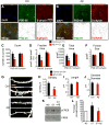
(A and B) At DIV 21 mES derived neurons were treated for 24 hours with DMSO control (Ctrl) or Aβ42 oligomer, subsequently fixed and co-stained for synaptic proteins PSD-95 (green) and synpatophysin (S-physin) (red). Nuclei are labeled with DAPI (blue). PSD-95 and S-physin particles were analyzed using Image J software. (C-F) Quantification of PSD-95 positive particles and S-physin positive particles using Image J software particle analysis. (G) DIV 21 ESN were treated with control or Aβ42 oligomer, fixed, diolistically labeled and imaged with confocal microscopy. (H-J) Spine density, length and dendrite diameter were quantified using Image J software. (K) Phosphorylation of CREB was detected with a pCREB specific antibody after no stimulation (NS) or NMDA stimulation (ST) of cultures with or without Aβ42 oligomer (Aβ) pretreatment. (L) Quantification of CREB and pCREB using infrared quantitative Western blot imaging system with SEM shown by error bars.
Figure 3
Figure 3. ESNs Harbor Machinery for APP Processing and Recapitulate Phenotype Associated with PS1 FAD
(A) APP and BACE1 expression in mES cells and ESNs at DIV 7, 14 and 21. (B) Components of the γ-secretatse, PS1, APH-1, NCT and PEN2 detected by Western blot analysis of the cell lysates using indicated antibodies. (C) Gradient centrifugation shows APP and BACE1 are enriched in the late endosome compartment with Syn6 and Rab11 but not at the top of the gradient with EEA1. (D) Western blot analysis of protein lysates from mES cell derived neurons infected with Lentivirus harboring human APPsw expressing APP and proteolytic fragents. Secreted APP fragments, sAPPβ and sAPPα, were immunoprecipitated from conditioned media and APP and CTFs were detected in cell lysates. (E-F) Aβ40 and Aβ42 were detected in conditioned medium using human specific ELISA and values were normalized to protein lysate concentration. (G) Three different FAD mutants of PS1 (ΔE9, M146V, and L286V) were introduced into mES cells which were subsequently subjected to directed differentiation into pyramidal neurons. A representative Western blot shows transgene expression in the neurons derived from these clonal mES cell lines using an anti-human PS1 specific antibody (PS1-NT). (H) PS FAD-expressing cells undergo normal directed differentiation into pyramidal neurons indicated by expression of neuronal proteins. (I) ELISA detection of Aβ42 and Aβ40 from conditioned media of ESNs stably expressing PS-FAD mutants infected at DIV 7 or 14 with APPsw Lentiviral particles resulted in enhanced ratio of Aβ42/Aβ40. Lentiviral gene transfer did not affect the viability of ESNs (data not shown).
Figure 4
Figure 4. Analysis of mES Cells and ESNs Isolated From Tg2576 AD Mouse Model
(A) Tg2576 mES-derived neurons identified by nuclear DAPI stain, express neuronal β-tubulinIII (TUJ-1) (green) and pyramidal cell protein EMX1 (red). Scale bar represents 40μm. (B) Western blot analysis of protein lysates from Tg2576 mES-derived neuronal culture harvested at indicated DIV express APP, cleaved APP fragments (APP-CTF) and neuronal proteins synaptophysin, CamKIIα and neuronal β-tubulinIII (TUJ-1). α-Actin is a loading control. (C) Cell lysates from DIV 7 Tg2576 ES-derived neurons treated with indicated compounds were subjected to Western blot detection with 6E10 (APP-FL), APP-CTmax (APP-CTF) and antibodies to indicated proteins. (D) Secreted sAPPα and APPβ were immunoprecipitated from conditioned medium after treatment of Tg2576 ES-derived neurons with the indicated compounds. (E, F) Secreted Aβ levels detected using human specific Aβ40 or 42 ELISA kit. Aβ40 and Aβ42 show characteristic sensitivity to BSI and GSI inhibition. IC50 values for the BACE1 inhibitor were 229.7nM and 28.1nM for Aβ40 and Aβ42 respectively. IC50 values for Compound E were 136.9pM and 102.2pM Aβ40 and Aβ42 respectively. The Z’ factors were 0.85 for Aβ40 and 0.5 for Aβ42 indicating an excellent assay for HTS.
Figure 5
Figure 5. Miniaturization and Optimization of 96 Well Platform for Mouse ESNs
(A) DIV 8 Tg2576 ES-derived neurons were plated at increasing density in a 96 well plate and subjected to a cell viability assay quantified in relative fluorescent units (RFU). (B) %CV and (C) Z’ were calculated with increasing number of wells. The minimum %CV and maximum Z’ are optimized at n=5 wells at a plating density of 1.5×10⁵ cells/cm2 (arrows in B and C).
Figure 6
Figure 6. Screening NIH Clinical Collection (NCC) for Aβ Biogenesis
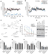
(A) DIV 7 Tg2576 ESNs plated in 96 well plate platform were treated with compounds from the NCC compound collection for 24 hours and Aβ40 was detected in conditioned media and is shown as % Control, DMSO treated wells. Negative control (Ctrl) was DMSO, while Positive Ctrl 1 was BACE inhibitor IV and Positive Ctrl 2 was Compound E. (B) The remaining cells were subjected to cell viability assay CellQuanti Blue. (C) Potent hits were obtained from an independent source and subjected to IC50 determination in the 96-well platform for Aβ40 biogenesis (Amiridine, 6.29 μM; Icariin, 1.412 μM; Phenelzine, 0.2834 μM; Progesterone, 0.1746 μM) determined by Prism analysis log(inhibitor) vs. normalized response curve fit with baseline level of Aβ40 subtracted. (D) Chemical structures of hit compounds(E) Representative Western blot analysis of APP processing determined using immunoprecipitation of secreted fragments α and β from condition media. Full length APP was determined in protein lysate using Western blot analysis with 6E10 antibody and CTFs were detected using CTMaxi antibody. Neuronal β-tubulin was detected using TUJ1. (F,G) Quantification of sAPPβ and sAPPα (H) Full length APP was quantified by Image J analysis and normalized to tubulin (TUJ1). (I) Quantification of Aβ40 after treatment with indicated compounds normalized to protein level. Shown are means and SEM (n=3).
Figure 7
Figure 7. Disparity In Pharmacological Response of Hit Compounds Between Tg2576 ESNs and Cell Lines
(A) Tg2576 ES cell derived neurons; Neuro2a cells stably overexpressing APPsw (N2a-APPsw); Chinese hamster ovary cells stably overexpressing wild type APP and PS1 with the ΔE9 mutation (CHO-APPPS1); and SH-SY5Y cells stably overexpressing GFP-BACE1 and HA-tagged wild type APP (BGWT8) were plated in 96 well plates and treated with hit compounds (10μM) for 6 hours after which media was harvested and tested for Aβ40 content which was normalized to DMSO treated cells for each type (% control). Absolute values for control (DMSO) treated cells are Tg2576 ESNs: 129±29.2 pg/ml; N2a-APPsw: 346±50.7 pg/ml; CHO-APPPS1: 62.5±.7 pg/ml; and BGWT8: 28.8±6.4 pg/ml.

References

    1. Abbott A. Neurologists strike gold in drug screen effort. Nature. 2002;417:109. - PubMed
    1. Barker RA. Stem cells and neurodegenerative diseases: where is it all going? Regen Med. 2012;7:26–31. - PubMed
    1. Bibel M, Richter J, Schrenk K, Tucker KL, Staiger V, Korte M, Goetz M, Barde YA. Differentiation of mouse embryonic stem cells into a defined neuronal lineage. Nat Neurosci. 2004;7:1003–1009. - PubMed
    1. Bibel M, Richter J, Lacroix E, Barde YA. Generation of a defined and uniform population of CNS progenitors and neurons from mouse embryonic stem cells. Nat Protoc. 2007;2:1034–1043. - PubMed
    1. Braginskaya FI, Zorina M, Pal’mina NP, Gaintseva VD, Burlakova EB, Selezneva ND, Kolykhalov IV, Gavrilova SI. Some blood biochemistry parameters during the cholinergic treatment of Alzheimer’s disease. Neurosci Behav Physiol. 2001;31:457–461. - PubMed

Publication types

Substances

LinkOut - more resources